Playlist
Show Playlist
Hide Playlist
Occipital Lobe

-
Slides 17 CerebralCortex BrainAndNervousSystem.pdf
-
Reference List Anatomy.pdf
-
Download Lecture Overview
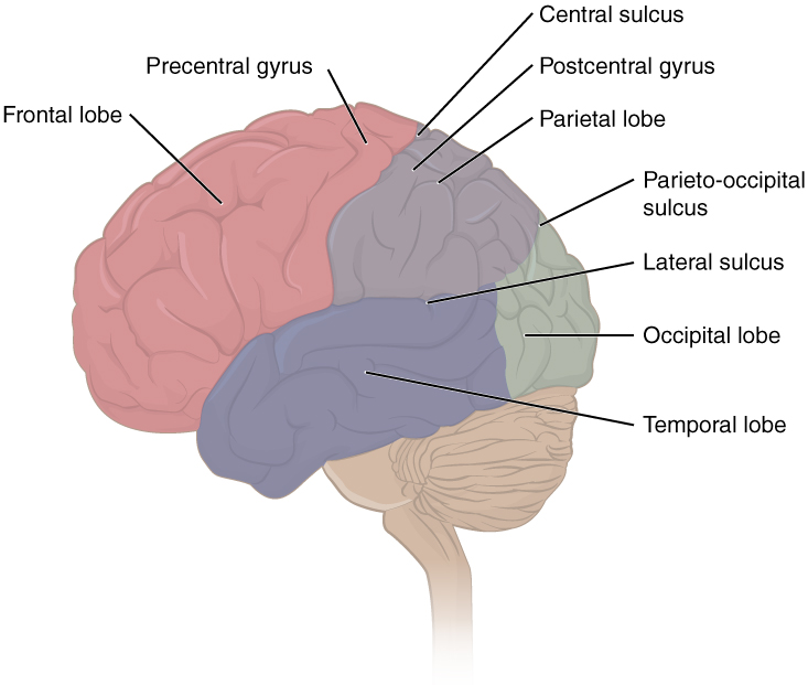
00:01 The next lobe is the occipital lobe. We see it in color on the posterior aspect of the illustration. 00:09 An area, a cortical area associated with the occipital lobe is that of the primary visual cortex. 00:17 That is labelled right in through here at the most posterior aspect of your occipital lobe. 00:23 This is where vision is perceived. A lesion to your primary visual cortex will result in contralateral homonymous hemianopsia. In addition, just lying anterior to your primary visual cortex, you will have a visual association cortex. You may see that right in through here. 00:50 This is processing various inputs that are related to vision and our ability to perceive and perceive it with respect to our surroundings. That is going to be form and color of the visual input. 01:11 This is the area that allows us to process motion. It allows us to understand the depth of our vision. It helps us to understand special relationships that are attached to visual processing. Bilateral lesions to the visual association cortex cause loss of color and loss of special relationships.
About the Lecture
The lecture Occipital Lobe by Craig Canby, PhD is from the course Cerebral Cortex.
Included Quiz Questions
Which of the following could be the result of a lesion in the primary visual cortex?
- Contralateral homonymous hemianopsia
- Ipsilateral homonymous hemianopsia
- Contralateral lower quadrant anopsia
- Hemianopsia with macular sparing
- Bitemporal hemianopsia
Which of the following is NOT a function of the visual association cortex?
- Control of conjugate lateral gaze
- Processing of motion
- Form and color of visual input
- Understanding spatial relationships
- Understanding depth of vision
Author of lecture Occipital Lobe

Craig Canby, PhD
Customer reviews
5,0 of 5 stars
| 5 Stars |
|
2 |
| 4 Stars |
|
0 |
| 3 Stars |
|
0 |
| 2 Stars |
|
0 |
| 1 Star |
|
0 |
Simple, clear and with clinical correlations. Thank you, Dr. Craig.
Dr Canby's fluent presentation is the first reason, also the simple connections between the anatomy and clinical correlation are very important to me.
