Playlist
Show Playlist
Hide Playlist
Non-Streptococcal Acute Glomerulonephritis

-
Slides Glomerulonephritis.pdf
-
Reference List Pathology.pdf
-
Download Lecture Overview
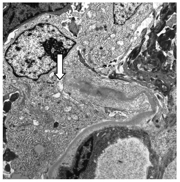
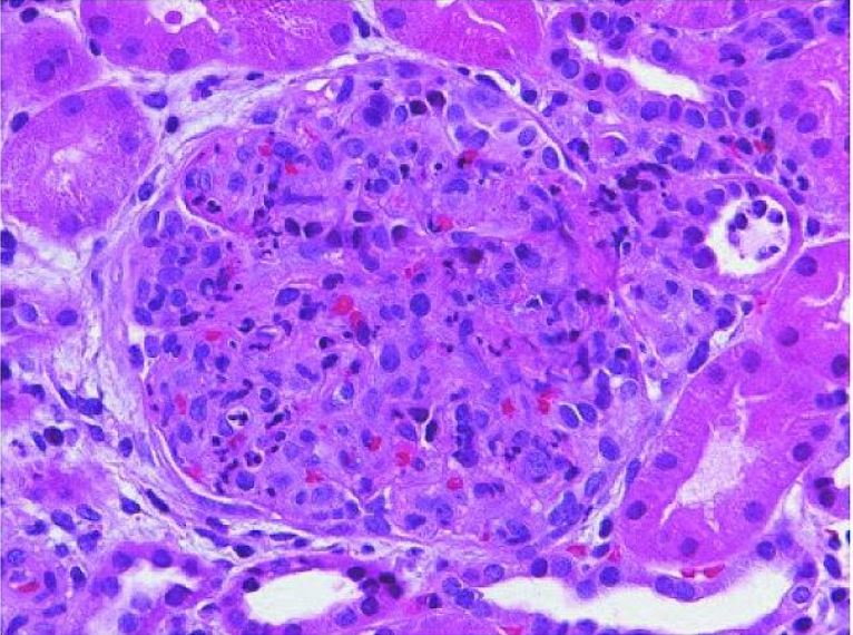
00:01 Now. Stop there. 00:03 And now let's move on to non streptococcal acute Glomerulonephritis. 00:08 Non streptococcal. 00:10 May be a result of non streptococcal infection such as staph endocarditis and pneumococcal, Meningococcemia, hepatitis B and C, mumps, HIV varicella You have a lot of viruses that are included here, EBV, and perhaps even parasitic. 00:25 So there are many other causes of Glomerulonephritis that would be rather infectious, but it'd be non streptococcus. 00:31 In this picture, what I'm showing you is the fallen. 00:36 What you're going to be paying attention to is the following order of ultra microscopic changes. 00:42 The first thing that you're always going to do with electron microscopy is for you to then identify the paved road, the smooth paved road is always going to be your basic membrane. 00:55 Your next step is then to measure or to identify the feet or the foot processes so what you will be finding when you find your foot processes is what cell? Good. 01:07 Take a look at visceral epithelial cells, which is highlighted here when you find such a deposit, which is dark density, which you see in electron microscopy here, those dark densities that are underneath the Visser epithelial cell then represent what going to deposit sub epithelial. 01:27 Next normal basic membrane has a light gray appearance. 01:30 That's the smooth paved road we've been referring to. 01:34 And then what we'll do next is, well take a look at them immunofluorescence. 01:38 The image of immunofluorescence upon such a deposit of immune complexes gives you a granular pattern. 01:44 Well, how can distinguish this for something that's linear? First and foremost, take a look at the history and then repeat repeat. 01:50 Repeat the number of images. 01:52 The one that you want to compare this to would be your linear. 01:55 This is not linear. 01:57 This is granular. 01:59 What did the granular mean to you? Oh, there's a mean, complex deposition. 02:03 It just happened to be that the immune complex deposition here for PSGN was sub epithelial. 02:10 Good. 02:11 Granular at some point, could also mean sub endothelium. 02:14 And if there a different history, well then it would be a different diagnosis.
About the Lecture
The lecture Non-Streptococcal Acute Glomerulonephritis by Carlo Raj, MD is from the course Glomerulonephritis.
Included Quiz Questions
Which of the following is correct regarding Group A streptococcal GN?
- Electron dense deposits are seen directly beneath the visceral epithelial cells.
- Electron dense deposits are seen directly beneath the parietal epithelial cells.
- Electron dense deposits are seen directly beneath the endothelial cells.
- A homogenous pattern is observed in immunofluorescence.
Author of lecture Non-Streptococcal Acute Glomerulonephritis

Carlo Raj, MD
Customer reviews
5,0 of 5 stars
| 5 Stars |
|
5 |
| 4 Stars |
|
0 |
| 3 Stars |
|
0 |
| 2 Stars |
|
0 |
| 1 Star |
|
0 |
