Playlist
Show Playlist
Hide Playlist
Neurovasculature of the Orbit
-
Slides Anatomy Eye.pdf
-
Download Lecture Overview
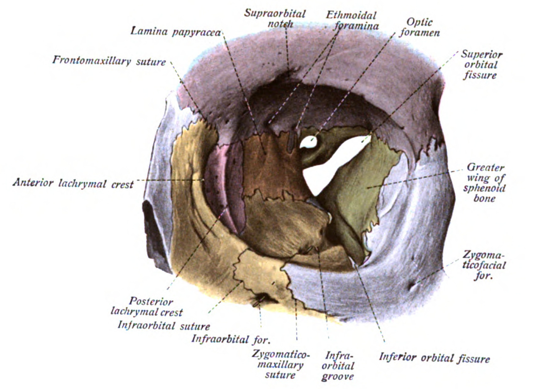
00:01 The arterial supply to the eye and the accessory structures of the orbit is derived from the ophthalmic artery, which is a branch of the internal carotid artery and enters the bony orbit through the optic canal along the optic nerve. 00:17 All other arteries we shall discuss now are branches of the ophthalmic artery. 00:22 The first branch is usually the central retinal artery that travels along and sometimes in the dermal sheath of the optic nerve to reach the globe of the eye. 00:31 Once it reaches the eye, divides into superior, inferior, nasal and temporal branches to supply the retina. 00:39 Blockage of blood flow through this artery manifests as a unilateral of blindness. 00:45 The next branch is the lacrimal artery which courses on the lateral aspect of the orbit to reach and supply the lacrimal gland. 00:54 Along the way, it also gives rise to the lateral palpebral arteries which supply the eyelid and conjunctiva. 01:01 And an anastomosis with the medial palpebral arteries as well as the zygomatic branches which travel in the zygomatical temporal and zygomatic facial prominent to reach the temporal fossa and the cheek and then they will announce the most with various arteries in that location. 01:19 Next branches are the long in short posterior ciliary arteries. 01:24 The long posterior ciliary arteries go to supply the iris in the ciliary body and the short posterior ciliary arteries go on to supply the choroid. 01:33 After this, a supraorbital artery takes root and travel severely through the supraorbital foramen to supply the forehead and the scalp. 01:43 Then the ophthalmic artery continues anteriorly on the medial side it gives a posterior and anterior ethmoidal arteries which enter the posterior and anterior ethmoidal canals respectively. 01:57 Before giving off terminal branches, the ophthalmic artery gives rise to the medial palpebral arteries that anastomosis with the lateral palpebral arteries to supply the eyelid. 02:09 Terminally the ophthalmic artery branches to give rise to the supra trochlear artery which travels superiorly to anastomosis with the supraorbital artery and supply the forehead and scalp and the dorsal nasal artery which supplies the nasal lacrimal sac. 02:26 Lastly, throughout his course, the ophthalmic artery gives rise to muscular branches which supply the extra ocular muscles and give rise to anterior ciliary arteries which also supply the iris. 02:40 Additionally, those structures which are located near the floor of the orbit such as the inferior rectus, inferior oblique and the nasal lacrimal sac can also derive their vascular supply from the infraorbital branch of the maxillary artery which enters the orbital cavity via the inferior orbital fissure. 03:00 The veins of the orbit usually follow the arteries in generally all drain into the cavernous sinus either directly or indirectly. 03:09 The major veins include: The superior ophthalmic vein, which receives the nasal dorsal, ethmoidal, supratrochlear, supraorbital, lacrimal, vorticose, as well as a central retinal veins and then travels through the superior orbital fissure to drain into the cavernous sinus. 03:32 And the inferior ophthalmic vein which receives additional vorticose vein and passes through the inferior orbital fissure to drain into the cavernous sinus. 03:43 Additionally, the inferior ophthalmic vein can drain into the pterygoid plexus via a communicating vein. 03:51 The innervation of the orbit is a separate topic of its own and should be discussed separately in relation to the remainder of the cranial nerves. 03:59 However, throughout this lecture, when discussing the walls and openings of the orbit, we have touched upon these terms. 04:05 Therefore, I'd like to provide a brief overview which will help us later on when we discuss the extra ocular muscles and their innervation. 04:13 The orbit receives both somatic, motor and sensory innervation from various branches of the second, third, fourth, fifth and six cranial nerves. 04:25 More specifically, the motor innervation, the extra ocular muscles comes from the ocular motor, trochlear, and abducens nerves which are the third, fourth and six cranial nerves respectively. 04:39 We will come back to these nerves in slightly more detail when we discuss the extra ocular muscles. 04:46 Somatic sensory innervation of the orbit is derived mainly from the branches of the ophthalmic nerve, which is itself the first branch of the trigeminal or the fifth cranial nerve. 04:56 These branches include: The lacrimal nerve which entrance The lacrimal gland, the skin of the upper eyelid and the conjunctiva. 05:05 The frontal nerve and its terminal supraorbital nerve, which innervates the frontal sinus conjunctiva scalp forehead, in upper eyelid, also the supratrochlear nerve which innervates the forehead, scalp and upper eyelid, and then the nasociliary nerves, which further branches out into the infra trochlear, ethmoidal and long ciliary nerves. 05:30 Additionally, the infraorbital nerve, a branch of the maxillary nerve, which is itself the second branch of the trigeminal nerve also provides somatic sensory innervation to the lower eyelid. 05:43 The autonomic innervation of the orbit comes mainly from postganglionic long and short ciliary nerves, which provides sympathetic and parasympathetic innervation to various structures in the orbit such as the iris, the lacrimal gland and ciliary muscles. 06:01 And lastly, the somatic innervation, the orbit comes from the optic or the second cranial nerve. 06:07 Since the optic nerve is a track of the brain. 06:10 It is surrounded by the three meningeal layers that cover the nervous system. 06:14 The subarachnoid space extends along the nerve to the point where it attaches to the posterior aspect of the eyeball. 06:21 In case of an increase in intracranial pressure, that pressure will also compress the optic nerve and its venous return via the retinal veins. 06:30 This results in edema of the optic disc.
About the Lecture
The lecture Neurovasculature of the Orbit by Craig Canby, PhD is from the course Head and Neck Anatomy with Dr. Canby.
Included Quiz Questions
Blockage of which artery leads to unilateral blindness?
- Central retinal artery
- Ophthalmic artery
- Lacrimal artery
- Lateral palpebral artery
- Long branches of the ciliary artery
Which vein communicates with the pterygoid plexus?
- Inferior ophthalmic vein
- Superior ophthalmic vein
- Cavernous sinus
- Central retinal vein
- Lacrimal vein
Which nerves provide the majority of the autonomic information to the orbit?
- Long and short ciliary nerves
- Optic nerve
- Infraorbital nerve
- Abducens nerve
- Oculomotor nerve
Customer reviews
5,0 of 5 stars
| 5 Stars |
|
5 |
| 4 Stars |
|
0 |
| 3 Stars |
|
0 |
| 2 Stars |
|
0 |
| 1 Star |
|
0 |


