Playlist
Show Playlist
Hide Playlist
Neuroglial Cells

-
Slides Cells and Basic Tissues-Nervous System.pdf
-
Reference List Histology.pdf
-
Download Lecture Overview
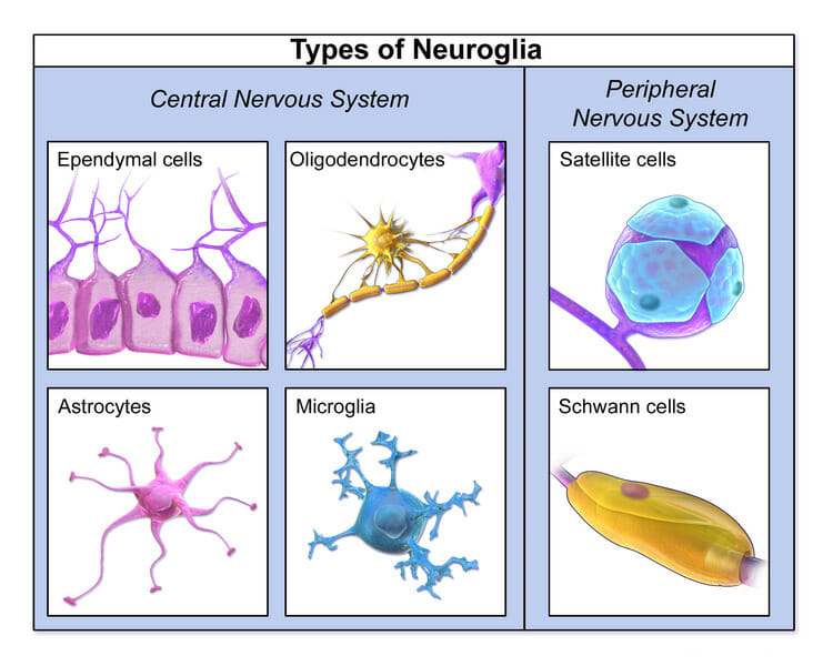
00:00 Well let us now look at supporting cells, the glial cells. Those cells that support the neurons and have a look at also their functions. We have learned already that ganglion cells are supported by satellite cells. Here you see ganglion cells in the top image. They are large cells, large neurons and all the little nuclei around them are the satellite cells. We have also learned in the previous couple of slides that nerve fibers are myelinated next done by the Schwann cell. Well there are other names for these supporting glial cells in other structures such as the endplate, the gut and in the retina. And we will come across those special glial cells when we look at those tissues in more detail. But there is a Schwann cell nucleus shown in this section and this Schwann cell nucleus is responsible for myelination in peripheral nerves. There is a satellite nucleus wrapping around or being part of the supporting cells around the big stained or big ganglion cell you see in the top image. Well let us look at the neuroglial cells in more detail. There are peripheral and central neuroglial cells. 01:34 The peripheral ones remember are the Schwann cells or the satellite cells that I have already pointed out to you. One of the most important central neuroglial cells is the oligodendrocyte. 01:49 It produces the myelin. So it is equivalent to the Schwann cell in the central nervous system. The Schwann cell is only responsible for myelination remember, in the peripheral nerve. Now they are quite easy to identify in sections. This happens to be a section through the gray matter of the spinal cord. Up at top left-hand side, you can see perhaps what is a ventral horn cell. It shrunk away from the tissue a bit and there is a whitish halo around it. On the far top right-hand corner, there is a space that happens to be the central canal of the spinal cord and I will refer to that in a moment. But have a look at the rest of the tissue. You can see some nuclei and they look different. 02:43 They are of different shape and they are of different size. And this is the best way of identifying the different glial cells, based on the shape of the nuclei and their size. 02:55 For instance, the oligodendrocyte is a very small round, dark stained nuclei and you can see that labeled here. One point about the oligodendrocyte, which makes it different to the Schwann cell apart from its location, is that oligodendrocyte actually can myelinate a number of axons whereas the Schwann cell cans only myelinate a single axon. Well, another very important glial cell is the astrocyte. It has a number of different jobs that I've listed there on the slide, but most importantly they form part of the blood brain barrier. When I talked about epithelia and I mentioned simple squamous epithelium forming a barrier, it does so in the brain. 03:51 The very thin epithelial cells that make up the lining of these capillaries in the brain, the endothelium, has very tight junctions and they prevent any materials going across into the neural tissues. It only allows glucose and oxygen to pass into that tissue and various other components that may be required. But these astrocytes actually help form that barrier by sending long projections up and encircling the capillary to almost be a double layer around these blood capillaries therefore creating a very important component of the blood-brain barrier. You can identify these cells because their nuclei are larger and often lot of staining as you see here. Well there is another important glial cell and that is the microglia cell. 04:54 They are like macrophages. They are phagocytic and you can see them easily in sections because they have an elonged sausage shaped nucleus. And finally the remaining type of glial cells that I want to bring to your attention are the ependymal cells. These line the fluid-filled cavities in the brain such as the ventricles and also the central space or central canal that I pointed out earlier. Well let us move on and look at the brain. Only certain parts
About the Lecture
The lecture Neuroglial Cells by Geoffrey Meyer, PhD is from the course Nerve Tissue.
Included Quiz Questions
Which of the following cells produces the myelin sheaths around neuronal axons in the peripheral nervous system?
- Schwann cells
- Myosatellite cells
- Muller cells
- Glial cells
- Oligodendrocytes
Which of the following cells plays a key role in the maintenance of the blood-brain barrier?
- Astrocytes
- Oligodendrocytes
- Muller cells
- Schwann cells
- Satellite cells
Which of the following cells in the central nervous system have elongated nuclei and mediate immune responses?
- Microglia
- Oligodendrocytes
- Ependymal cells
- Astrocytes
- Muller cells
Which of the following cells have phagocytic activities in the central nervous system?
- Microglia
- Oligodendrocytes
- Astrocytes
- Ependymal cells
- Satellite cells
Author of lecture Neuroglial Cells

Geoffrey Meyer, PhD
Customer reviews
5,0 of 5 stars
| 5 Stars |
|
1 |
| 4 Stars |
|
0 |
| 3 Stars |
|
0 |
| 2 Stars |
|
0 |
| 1 Star |
|
0 |
Short and to the point, good professor, and good test prep questions
