Playlist
Show Playlist
Hide Playlist
Names of Ducts of Glands

-
Slides 02 Types of Tissues Meyer.pdf
-
Reference List Histology.pdf
-
Download Lecture Overview
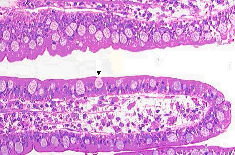
00:00 Let's look at how ducts are named in glands. Well, they can be named in a number of different ways. Firstly, you can name them according to the location you see these ducts. I mentioned a large compound gland earlier and that large compound gland is divided by connective tissue into large lobes and then smaller lobules. Well, histologically you have done often sections through ducts that are in, between different lobes, interlobular ducts. You only really see these small ducts. And those small ducts when they are located between a lobule is called an interlobular duct and within the lobule, it is called an intralobular duct. 00:57 You need to be able to identify these different ducts when you see sections through some of the glands. Often they are classified according to, not just say structure, but also their function. 01:11 They are called excretory duct if their job is only to convey the secretory product and most of the large ducts, the interlobular ducts are excretory ducts because they have no other function than carrying the secretional product to the location where the secretory product is going to be active. But a lot of secretory ducts are involved with changing the compounds, the components of the secretory products. They change the compounds, the components because they are involved with transport of material into the lumen and the absorption of material from the lumen. So they will have a secretory function just as much as the secretory cells do, absorption of material or secretion of material. And in this case, you will see in glands often ducts we termed striated ducts or intercalated ducts particularly in serous secreting or protein secreting glands. Remember these striated ducts and intercalated ducts are really involved with modifying the secretion product. Sometimes we name ducts according to where they are located. For instance, the ductus deferens in the male reproductive system sometimes they are named after the individuals who first described them. Well let's have a look at a couple of images illustrating certain ducts. Here is an interlobular duct. 02:57 Notice the secretory acini or the secretory units on the right hand frame of the slide, secretory units are serous because they are rather dark staining, pink staining. There are the granules inside the cells of the apex of the cell, the protein granules. But look at the duct, it is seperated from the secretory units by a fair of connective tissue, so that indicates that connective tissue is dividing the gland into different lobules. Down below that structure that has a reddish color in the middle happens to be a blood vessel. Normally, as I said earlier, blood vessels and ducts accompany these connective tissue septa seperating lobules from each other. Now have a look at this section. It is taken through a serous secreting gland. 04:00 What dominates are the white circular spaces you see. They happen to be fat cells or adipocytes, and similar to other mucous secreting cells I explained earlier, the content of these cells is lost during processing. Have a look at the left hand side of this picture and see if you can make out intralobular ducts. They have a circular shaped lumen, and if cut nice transversely, then they have a nice cuboidal epithelium. This is a high magnification again through a protein secreting gland showing striated ducts. Again, look at the tube on the left hand side of the image. It is a striated duct, but have a look at the luminal area and then move your eyes and look at the basal area of this epithelium, and notice that there are these little pinkish or reddish stripes running along the basal aspect of this epithelium. 05:16 That is why they are called striated ducts. And those striations represent firstly the arrangement of mitochondria in columns along the basal portion of the cell and these mitochondria are arranged wiithin many many basal folds of the epithelial cell. And these mitochondria are there to provide the energy for active transport because striated ducts are very important in protein secreting glands because they modify the secretory product. 05:53 So these mitochrondia provide the energy for active transport, to move material from the lumen into the underlying connective tissue. And the basal folding is a way of increasing the surface area to create more space for these transport channels and also to house many many more transport proteins. Now have a look this slide very carefully. It again is a serous or a protein secreting gland. Pickout the secretory units or secretory acini and acinous is a grape, berry like structure. It is a cluster of cells. Let's see if you can find an intercalated duct. They are very hard to see. They are very, very very small in dimension and very short and really only obvious in protein secreting glands, not mucous secreting glands because in mucous secreting glands, the secretion product is not modified. But in protein secreting glands as I have indicated earlier, it is modified. So these ducts are also quite prominent when you see sections of protein secretory glands. Well have you found that? There it is. 07:22 A little tiny white spacious lumen and if you look very carefully you see a cluster of cuboidal type epithelial cells surrounding or making that lumen. That is an intercalated duct. Well, ducts have many functions, which I have explained as we have gone through this lecture. 07:50 One is just to convey secretions and we call it an excretory duct. Sometimes the duct needs to modify the secretion product of the gland and we will see examples of that when we look at sweat glands in another lecture on skin and also when we look at the salivary glands. 08:10 Sometimes ducts store secretion. When we look at the prostate gland in the male reproductive system and also the mammary gland, we will learn that when the secretion product is produced by the cell, it is often stored in the duct system until it is used or wanted.
About the Lecture
The lecture Names of Ducts of Glands by Geoffrey Meyer, PhD is from the course Epithelial Tissue.
Included Quiz Questions
In exocrine glands, which of the following ducts is lined by cells with basal infoldings of their cell membranes, associated with many mitochondria and transport channels?
- Striated duct
- Excretory duct
- Intercalated duct
- Intralobular duct
- Interlobular duct
Which of the following connects the acinus to a striated duct in an exocrine gland?
- Intercalated duct
- Intralobular duct
- Interlobular duct
- Excretory duct
In exocrine glands, which of the following ducts, along with striated ducts, function to modify salivary fluid?
- Intercalated ducts
- Stensen’s ducts
- Interlobular ducts
- Alveolar ducts
- Tubular ducts
Which of the following statements about the ducts in glands is INCORRECT?
- Striated ducts cannot modify the secretions.
- Intercalated ducts are often difficult to identify in mucus-secreting glands.
- Intercalated ducts are involved in the modification of secretory products.
- Interlobular ducts are found in between gland lobules.
Author of lecture Names of Ducts of Glands

Geoffrey Meyer, PhD
Customer reviews
5,0 of 5 stars
| 5 Stars |
|
5 |
| 4 Stars |
|
0 |
| 3 Stars |
|
0 |
| 2 Stars |
|
0 |
| 1 Star |
|
0 |
