Playlist
Show Playlist
Hide Playlist
Myocyte Conduction Pathway and Atrioventricular Block (AV) – Heart Rate and Electricity
-
Slides Heart Rate and Electricity.pdf
-
Download Lecture Overview
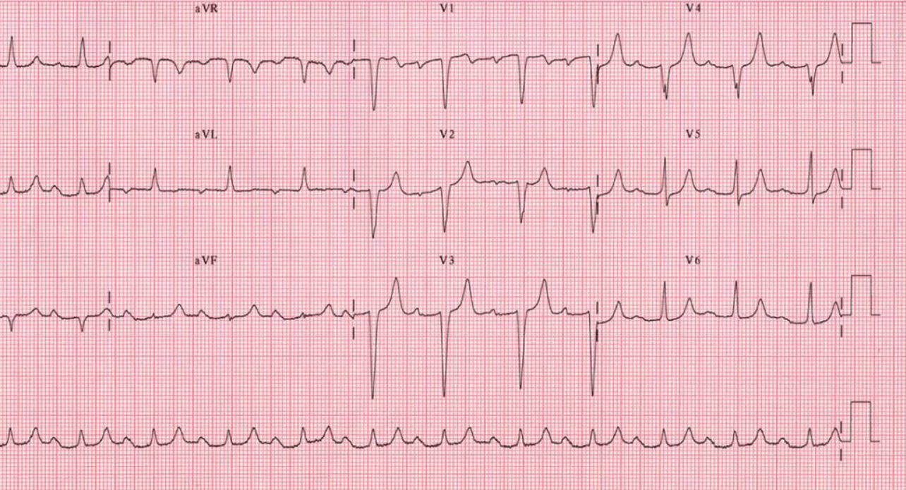
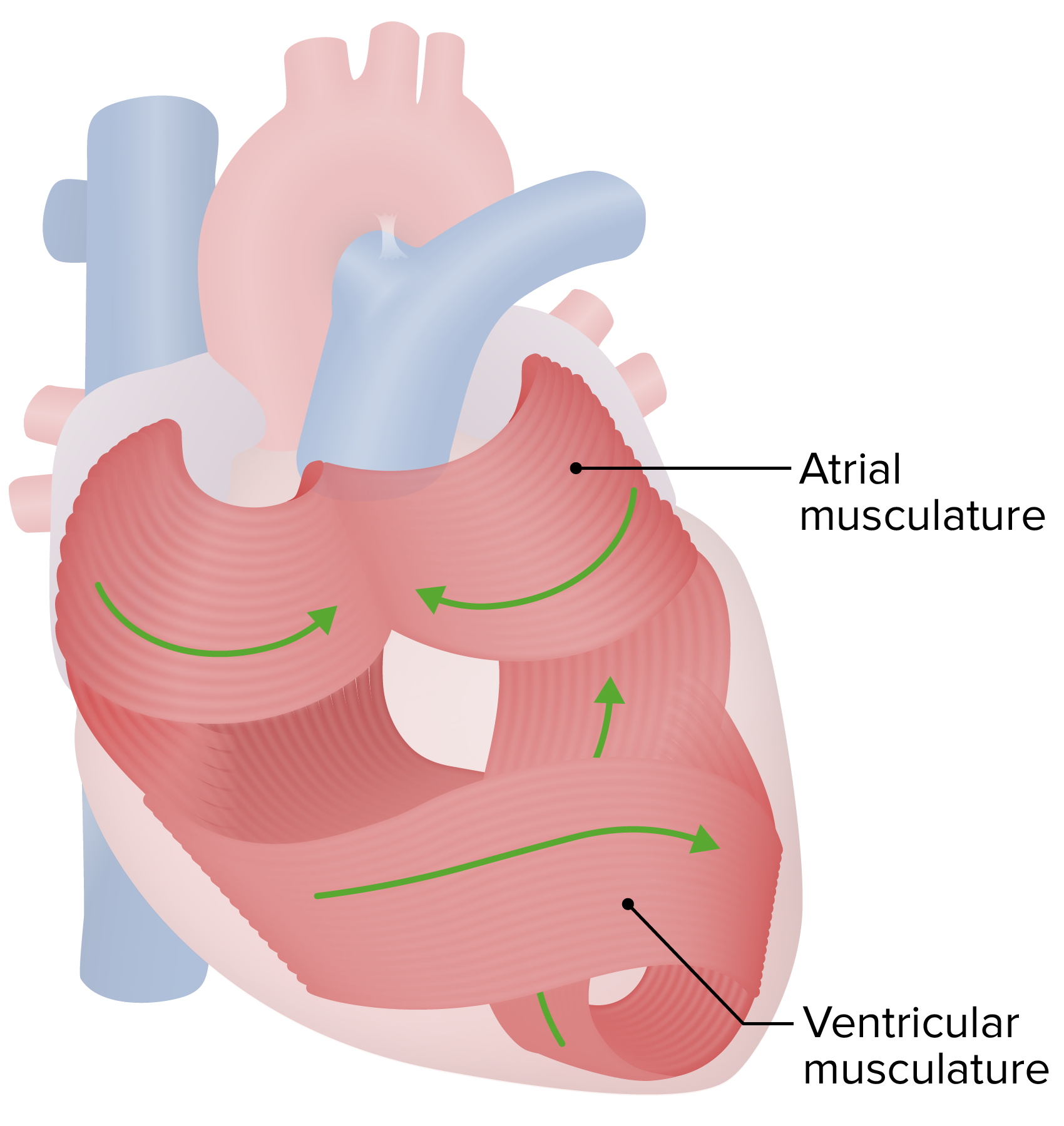
00:01 Let's talk through how you get a signal from the SA node, all the way down to these cardiac myocytes. 00:10 There are some specific pathways, by which you’re going to go through. 00:15 So, you start at the SA node, you go to the AV node, you go down the Purkinje fibers and that is how this process works. 00:24 But I'll tell you something. 00:26 Some of the places along this conduction pathway are faster than others. 00:31 So, let's start with the fastest stuff, right? The fastest thing we have are Purkinje fibers. 00:38 Purkinje fibers are really quick conductors. 00:41 So, as so soon as they get electricity, boom, they move it right on. 00:46 Right behind that is the right and left bundle branches. 00:49 So, those are the areas right along the septum. 00:52 They also conduct electricity very rapidly. 00:57 The ones that conduct it a little bit are in the atrial muscle and ventricular muscle. 01:04 This is kind of a moderate level of electrical conduction in terms of speed. 01:11 What’s the slowest – what is the slowest process in this whole thing? The weakest link. 01:17 It is the slow portion and that is the AV node. 01:22 So, the AV node slows things down. 01:26 It is a slow conductor, while the other things like Purkinje fibers move really quick. 01:33 So, it just depends upon which area you are in the heart, of which of these particular conduction velocities will allow for the process to happen. 01:45 If you think about it, it makes sense that these will have different rates. 01:50 Why? Well, first, you need to signal the top portion of the heart to contract, but you need to wait until the ventricles fill up before you contract them. 02:02 And that's one of the primary purposes of the slow AV node that allows it to slow down, so you have enough time to fill the ventricles. 02:13 Once the ventricles are full, then you can contract them optimally. 02:18 But you can't have that process, you couldn’t depolarize the whole heart at the same speed or you wouldn't get this pumping ability. 02:27 Once you have information sent down the bundle branches to Purkinje fibers, that is important to happen very quickly because you want to make sure that they contract as a unit. 02:39 So, these conduction velocity speeds make a lot of physiological sense and that is why we go through them in this way, so you have a good feel for how the conduction process works with the mechanics and how this process works to be able to pump blood throughout the body. 03:00 So, there are a couple of factors that affect the conduction velocity. 03:05 The first is sympathetic stimulation. 03:08 So, this fight or flight response or this fight or aggressive conflict mediation response allows for there to be faster conduction of current. 03:18 We call this positive dromotropy. 03:22 So, dromotropy is speed of conduction. 03:27 What slows down the speed of conduction? The parasympathetic nervous system. 03:32 And these are done through acetylcholine. 03:34 This decreases the conduction through places like the AV node, and we call that negative dromotropy. 03:43 So, we have chronotropy, is heart rate. 03:45 Dromotropy is speed of conduction. 03:51 Now, anytime we talk about pathology, there are places where this whole process could go awry, right? It seems like that anytime we talk about something how neat it is, how fast it is, how cool it is, we go, okay there's a place when – there’s a time when it is going to go wrong. 04:10 And there, of course, is in the conduction pathway as well. 04:14 The AV node is one area of pathology a lot of problems happen with the AV node, in that maybe it slows conduction too much and doesn't let it move through very rapidly. 04:27 There also can be blocks in places like the Bundle of His. 04:33 There can be also blocks in places like the bundle branches, the right or the left. 04:38 And each one will look a little bit different on electrocardiogram. 04:42 There also can be some other blocks downstream, but they’re a little less likely, and so what we do is we focus on the bundle branches and the AVs because they are the most important. 04:54 Because we cover the most important stuff here. 04:59 So, let's walk through a couple of the AV blocks. 05:03 So, what do we mean by AV block? It doesn’t always mean a block. 05:07 It doesn't mean that it doesn't travel through at all. 05:10 What we’re talking about here is that it slows it down, maybe it delays it, or maybe it doesn't let it go through. 05:18 But it doesn't mean that it's fully blocking it until you get to a type III block. 05:23 So, what is a type I block? It delays the conduction through the AV node. 05:28 So we just have here – think of it like a gate. 05:31 You know, you've been on the interstate and you've been on a large highway, in which all at once, you have a tollgate out and it slows everybody down. 05:42 And it’s these kind of gates that don't stop movement of traffic, but they slow that movement of traffic down. 05:51 So, the interesting thing about type I AV blocks is that the rate is lower, but you still have a normal sinus rhythm. 06:00 What do we mean by a sinus rhythm? It has a P wave, a QRS complex, and a T wave. 06:08 If we move to a more serious block, like a type II block, here is where some action potentials don't make it through. 06:17 Many of them are slowed down, but some don't make it through. 06:21 There are a couple of different types of AV blocks that are type II. 06:26 There's a type I and a type II. 06:28 They usually refer to this as Mobitz type I and Mobitz type II. 06:32 But these can be different ways in which you slow rates through the AV node and this results in a bradycardia. 06:41 Heart rate below 60. 06:44 So, this is a time when a slow heart rate is not necessarily in an endurance athlete. 06:51 It's in pathology in someone who can't have their heart beat fast enough because they have one of these AV blocks. 07:00 The most serious problem here are AV blocks that are AV type 3. 07:05 These do not allow for the propagation of an action potential from the SA node to the Bundle of His. 07:13 So, this is even a more problematic condition. 07:17 You will no longer have a sinus rhythm. 07:19 And you'll have a severe ventricular bradycardia. 07:23 In fact, the ECG will look much different because there will be some P waves, which is atrial depolarization, with no captured QRS complexes. 07:34 We’ll review this subsequently. 07:37 We have some time denoted just to look at what an ECG looks like because we know it's hard for you, so we want to make sure you got it. 07:45 So, we spend some extra time on that in a future time.
About the Lecture
The lecture Myocyte Conduction Pathway and Atrioventricular Block (AV) – Heart Rate and Electricity by Thad Wilson, PhD is from the course Cardiac Physiology. It contains the following chapters:
- Myocyte Conduction Pathways
- Possible Sites of Conduction Blocks
Included Quiz Questions
Which of the below elements of the cardiac electrical pathway are the fastest conductors?
- Purkinje fibers
- Atrial muscle
- Bundle branches
- AV node
- Ventricular muscle fibers
Which of the following elements of the cardiac electrical pathway are the slowest conductors?
- AV node
- Ventricular muscle
- Purkinje fibers
- Atrial muscle
- Bundle branches
Which of the following terms is associated with the activation of the heart by the sympathetic nervous system?
- Positive dromotropy
- Positive lusitropy
- Negative inotropy
- Negative chronotropy
- Hypertrophy
Which of the following conditions results in complete dissociation of atrial and ventricular activity?
- Third-degree AV block
- First-degree AV block
- Type I second-degree AV block
- Atrial defibrillation
- Type 2 second-degree AV block
Customer reviews
5,0 of 5 stars
| 5 Stars |
|
4 |
| 4 Stars |
|
0 |
| 3 Stars |
|
0 |
| 2 Stars |
|
0 |
| 1 Star |
|
0 |
Thanks for this easy explanation even people who never studied in Medical schools will still understand everything you explain
It was Very clear, had good retoric and the picures were easy to remeber
I really appreciate his enthusiasm and clarity. He's cheesy, but that's what makes his presentations a little more interesting.
I've been watching through and writing notes your videos til this one (thank you so much for making this easy and understandable while staying on a detailed level that I need for my exams) BUT there's something that wasn't that clear to me. What did you mean on the slide about type 2, where you mentioned Mobitz? Sentence nr2 of the 3 didn't make much sense to me.


