Playlist
Show Playlist
Hide Playlist
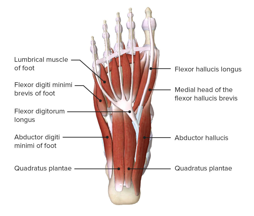
Muscles of Sole of Foot: Layer 2 – Anatomy of the Foot

-
Slides 07 LowerLimbAnatomy Pickering.pdf
-
Download Lecture Overview
00:00 also flex the fifth digit. So if we now move on to layer 2, we can see that we’ve had flexor digitorum brevis removed. Abductor digiti minimi is still present, and abductor hallucis is still present. But really, layer 2 is concerned with this arrangement of muscles in this central compartment. So we have quadratus plantae and we have lumbricals. And these are really important. If we look at quadratus plantae, we can see that’s running forwards and it’s actually inserting into the tendon of flexor digitorum longus. So a contraction of this muscle is going to support flexor digitorum longus. It may as well straighten the tendons so the line of pull is more efficient. Then we have the lumbricals, and we have four of these lumbricals. And they’re similar to lumbricals we observed in the hand. 00:56 They’re originating from the tendons of flexor digitorum longus as well. So if we look at quadratus plantae, we see it comes from the medial and lateral aspect of the calcaneus, and it inserts into the lateral margin of the tendons of flexor digitorum longus. It’s supplied by the lateral plantar nerve. And as I mentioned, this muscle is important in flexing or assisting the flexion of digits 2 to 5 with flexor digitorum longus. The lumbricals, just like the lumbricals in the hand, are coming from the tendons of flexor digitorum longus. And they insert into the medial aspect of digits 2 to 5. So they pass from the tendons of FDL to the medial aspect of the digits 2 to 5. Their nerve supply is different. The medial lumbricals are supplied by the medial plantar nerve, and the two lateral lumbricals are supplied by the lateral plantar nerve. 01:58 So the medial two lumbricals are supplied by the medial plantar nerve and the lateral two lumbricals by the lateral plantar nerve. These muscles have an important function. 02:08 They are important in flexing the proximal phalanges and extending the middle and distal phalanges of digits 2 to 5. So it’s very similar function to the lumbricals in the hand. Now let’s move on to layer 3, which contains a few more muscles. We’re going
About the Lecture
The lecture Muscles of Sole of Foot: Layer 2 – Anatomy of the Foot by James Pickering, PhD is from the course Lower Limb Anatomy [Archive].
Included Quiz Questions
Which muscle does the quadratus plantae assist with flexion of digits 2 - 5?
- Flexor digitorum longus
- Flexor hallucis longus
- Flexor digitorum brevis
- Abductor digiti minimi
- Flexor accessories
Author of lecture Muscles of Sole of Foot: Layer 2 – Anatomy of the Foot

James Pickering, PhD
Customer reviews
5,0 of 5 stars
| 5 Stars |
|
5 |
| 4 Stars |
|
0 |
| 3 Stars |
|
0 |
| 2 Stars |
|
0 |
| 1 Star |
|
0 |
