Playlist
Show Playlist
Hide Playlist
Mucosa and Serosa

-
Slides 02 Types of Tissues Meyer.pdf
-
Reference List Histology.pdf
-
Download Lecture Overview
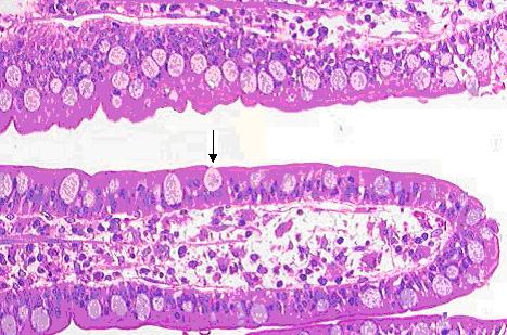
00:00 For now, let me explain the term mucosa and also serosa. Remember when I showed you the image of the stomach mucosa, I asked you to remember that word, that term, mucosa, because in subsequent lectures, I am going to use the term mucosa and also serosa quite often. 00:22 So it is very important to understand what I mean about these terms and they relate to glandular epithelium. They relate to the glands that are just being describing, the exocrine glands. Well you are familiar with this section on the left hand side. It is a section through the intestinal villus. And it shows the epithelial surface sitting on the lamina propria. 00:56 Remember the intestinal villus is a finger like projection that protrudes into the lumen of the intestine. 01:06 Well, it is actually an example of a mucosa. A mucosa lines cavities that are connected to the outside of the body, such as the intestine. We will see mucosas also in the respiratory passages, and in the urogenital system. Again, epithelial surfaces that line cavities exposed to the outside of the body. So when we look at an endothelium later on or the epithelium lining a blood vessel, we do not call them mucosa. Why? Because blood vessels are not connected to the outside of the body. On the right hand side, this is a very low magnification of a section through a part of the small intestine and those long extended processes are actually gross anatomical components of the small intestine and where the label of mucosa is shows these villi under very very low magnification. But remember, the villus, the epithelium and the underlying lamina propria plus sometimes a little bit of a little muscle layer you might see there is called the mucosa. It is facing in towards the lumen. It is a cavity of a tube that is connected to the outside of the body, which is what the gut is. Here is another example of the mucosa. This is a stomach mucosa that I asked you to recall earlier on the lecture. 02:53 So now you know what the mucosa means. It is labeled here under low magnification on the right hand side, underneath it's supported by what we call a submucosa, and I will talk about that term later on when we look at the description of the structure of the gut wall. 03:16 But again it is just to emphasize that the mucosa is the surface epithelium and supporting connective tissue or lamina propria, which we do not see a lot here because the mucosa is dominanted by secretory cells. But it's facing the lumen and it is in a tube that is exposed to the outside of the body through the esophagus and the other parts of the gut. Well let's look at the serosa. A serosa is an epithelial surface that lines cavities through the body that are closed. They are not exposed to the exterior of the body. Cavity such as the peritoneal cavity, the thoracic cavity, the pleural cavity, the pericardial cavity. All those surfaces are lined by what we called a serosa, a very thin simple squamous epithelium that is found on the surfaces of these cavities. On the right hand side, it is actually part of the large intestine or the colon. If you look inside of the lumen and you see the many folds stained very purple in this section, that is the mucosal surface in contact with the lumen. 04:41 It is a mucosa because it is lining the cavity of the gut here, which is exposed to the outside of the body, but on the other side of the gut wall, the serosa is exposed to the abdominal cavity, the peritoneal cavity. So that is what is called a serosa. 05:02 When I lectured about epithelium, I mentioned that one function of simple squamous epithelium is to act as a lubricating surface. Well, often in these cavities, there is a serous secretion or watery secretion that helps lubricate these cavities and helps create a friction free environment because often these cavitiies have contents moving all time. The lungs increasing and decreasing during breathing, the pumping of the heart inside the pericardial cavity requires a friction free environment, a surface that is lubricated by the serous secreting membranes or cells. Well in summary then, make sure you understand what a gland is. It is a secretory unit, derived from epithelial cells. Make sure you are aware and can identify the difference between serous and mucous secreting acini, or little clusters of secretory cells and mucous and serous secreting glands. Know the different ways in which the secretion product is delivered to the luminal surface and know how glands are named, simpler compound. And how they're classified by the way they appearm, are they unicellular or multicellular. 06:33 And finally know and appreciate that ducts are very important in some glands because they can modify the products, in other glands, they merely convey the product to the surface or to where the secretory product is active. So, I hope you enjoyed this account of exocrine glands. I hope you now know something about these glands and I hope you also look forward to learning in a later lecture about endocrine glands and also paracrine glands. 07:07 Thank you very much for listening.
About the Lecture
The lecture Mucosa and Serosa by Geoffrey Meyer, PhD is from the course Epithelial Tissue.
Included Quiz Questions
Which of the following statements regarding mucosa is most accurate?
- It lines cavities in the body that lead to the outside of the body.
- It is the layer beneath the epithelium.
- It is located beneath the lamina propria and muscularis mucosa.
- It lines the pleural and pericardial cavities.
- It does not line the mouth or the nose.
Which of the following structures is NOT lined by mucosa?
- Artery
- Stomach
- Intestine
- Tongue
- Anal canal
Which of the following is lined by serosa?
- Pericardial cavity
- Vagina
- Trachea
- Nasal passages
- Uterus
Author of lecture Mucosa and Serosa

Geoffrey Meyer, PhD
Customer reviews
5,0 of 5 stars
| 5 Stars |
|
1 |
| 4 Stars |
|
0 |
| 3 Stars |
|
0 |
| 2 Stars |
|
0 |
| 1 Star |
|
0 |
Magnificent! I love it! Especially the explanation of serosa? Nothing to say I have gentleman!!
