Playlist
Show Playlist
Hide Playlist
Midbrain
-
Slides 6 BrainStem BrainAndNervousSystem.pdf
-
Reference List Anatomy.pdf
-
Download Lecture Overview
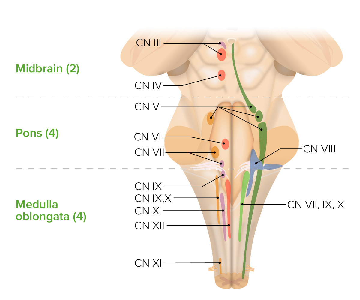
00:00 Some features of the midbrain for you to keep in mind as well as some of the functions. 00:06 Superior colliculus is shown here. Again, this will orient the head and the eyes toward a visual stimulus. 00:15 The red nucleus is shown in through here as well as over here. The red nucleus is receiving motor input from two sources, cortex and cerebellum. Then this will help to facilitate flexor movements in the contralateral upper extremity or limb. Some other features of the midbrain that are prominent here would be the presence of the substantia nigra, this black staining substance, black body is the little translation here or black substance. This is a member of the basal ganglia. There’s depigmentation of the substantia nigra due to a loss of dopaminergic neurons. This loss of dopaminergic neurons would characterize Parkinson’s disease. We can also see the anterior portion of the cerebral peduncle here. 01:18 This is the crus cerebri that we mentioned earlier. Additional features of the midbrain would include the cranial nerve III nucleus and the nerve. The nucleus is this slender structure here and here. 01:37 Then in this sectional region, you can see the cranial nerve III actually coming from the midbrain. 01:46 Also, you have the cerebral aqueduct that is shown here. This is a communication between the third ventricle and the fourth ventricle.
About the Lecture
The lecture Midbrain by Craig Canby, PhD is from the course Brain Stem. It contains the following chapters:
- Midbrain
- Arterial Supply
Included Quiz Questions
Which of the following statements about the red nucleus is most accurate?
- It facilitates flexor movements in the contralateral upper limb.
- It receives sensory inputs from the spinal cord.
- It has major effects on the lower limbs.
- It facilitates extensor movements in the ipsilateral upper limbs.
- It sends output signals to the cerebellum.
Customer reviews
5,0 of 5 stars
| 5 Stars |
|
5 |
| 4 Stars |
|
0 |
| 3 Stars |
|
0 |
| 2 Stars |
|
0 |
| 1 Star |
|
0 |


