Playlist
Show Playlist
Hide Playlist
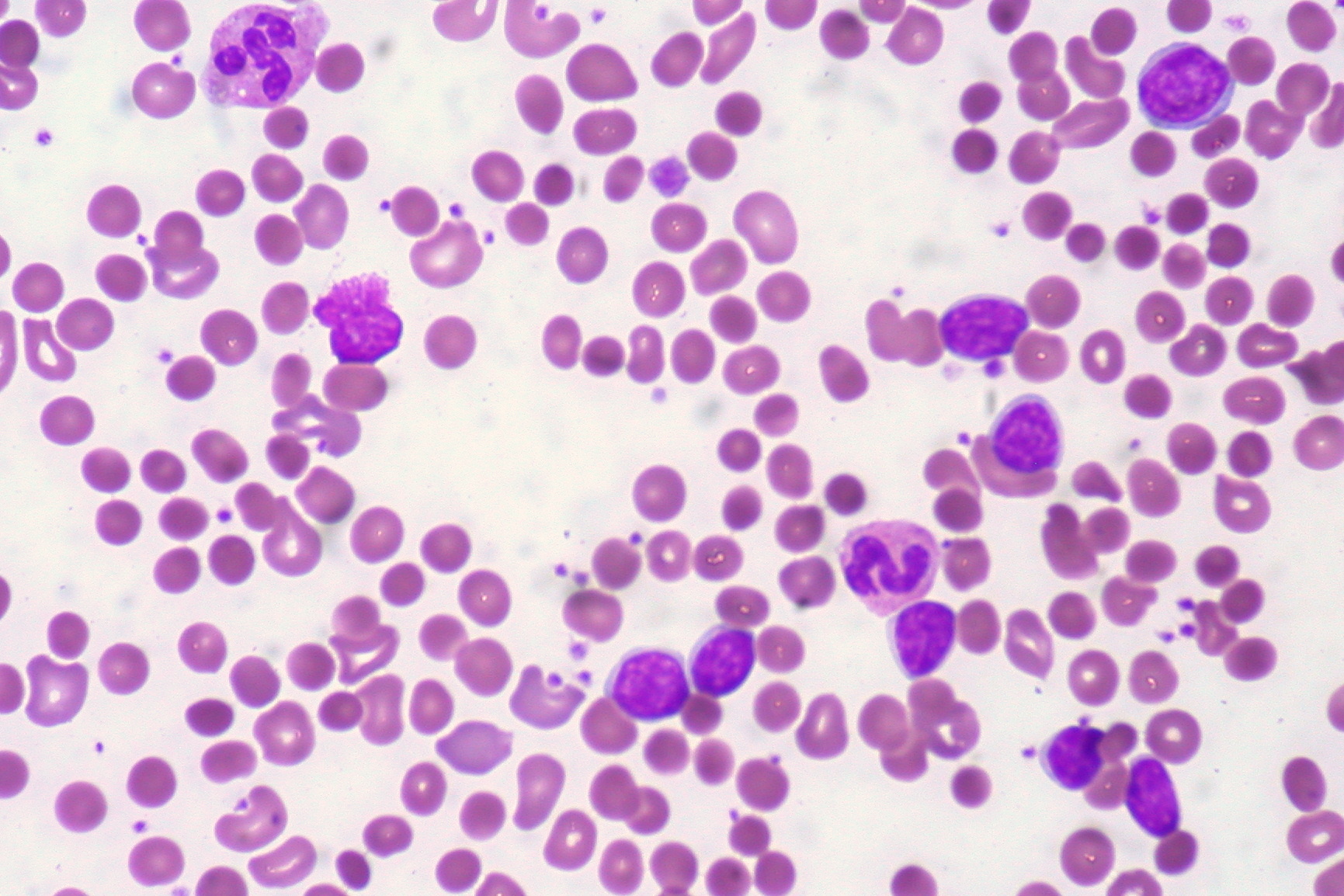
Microangiopathic Hemolytic Anemia: Pathogenesis and Morphology

-
Slides Microangiopathic Hemolytic Anemia.pdf
-
Reference List Pathology.pdf
-
Download Lecture Overview
00:00 Now, TTP and HUS, TTP is to an adult as HUS is to a child. 00:06 In these conditions where the thrombi are present, what happens to your RBC? It get’s sheared. 00:12 By sheared, what that means they’re not called sheared cells. 00:17 Let me make that clear. 00:18 However, you can use the word sheared, meaning that it gets broken up or fragmented and you can use the term to then call them clinically on a peripheral blood smear as schistocyte. 00:31 Now, be careful. 00:32 Do not confuse these with bite cells. 00:34 Bite cells are something that we saw with G6PD deficiency. 00:38 And ultimately, maybe, you want to wear this schistocyte on your head. 00:43 What? A helmet cell is sometimes is what it’s referred as its nickname. 00:50 Schistocytes is what it’s called clinically and technically on your peripheral blood smear. 00:55 But do not confuse them with sickle cells, do not confuse them with bite cells. 01:00 Look for the history, history, history. 01:02 If your patient has been exposed to amniotic fluid emboli, DIC, without a doubt, that picture there is going to be a schistocyte of an RBC, not a sickle cell, you see what I’m saying? So go back to the history so that you are clear about what kind of RBC you’re looking at. 01:18 Do that always because you can never fully guarantee. 01:22 Now, what you find in your peripheral blood smear is only for that disease. 01:26 It gives you an approximation, I’ll give you that. 01:29 But you must confirm your finding by looking at the history and making sure that you’re able to confirm it.
About the Lecture
The lecture Microangiopathic Hemolytic Anemia: Pathogenesis and Morphology by Carlo Raj, MD is from the course Hemolytic Anemia – Red Blood Cell Pathology (RBC).
Included Quiz Questions
What type of abnormal RBCs are likely to be seen in the peripheral blood smear of a patient suspected to have disseminated intravascular coagulation?
- Helmet cells
- Burr cells
- Bite cells
- Teardrop cells
- Sickle cells
Author of lecture Microangiopathic Hemolytic Anemia: Pathogenesis and Morphology

Carlo Raj, MD
Customer reviews
5,0 of 5 stars
| 5 Stars |
|
5 |
| 4 Stars |
|
0 |
| 3 Stars |
|
0 |
| 2 Stars |
|
0 |
| 1 Star |
|
0 |
