Playlist
Show Playlist
Hide Playlist
Micro- and Macroangiopathic Hemolytic Anemia esp. schistocytes

-
Slides Microangiopathic Hemolytic Anemia.pdf
-
Reference List Pathology.pdf
-
Download Lecture Overview
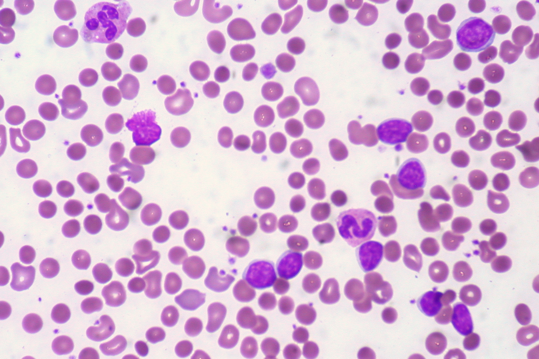
00:00 What we have here, schistocytes, is a picture in which exactly that. 00:04 It looks like you have cells in there in which you could wear on your head. 00:07 In other words, referring to helmet cells and classically called schistocytes. 00:12 What we're seeing here would be a aortic valve, which has undergone massive calcification, dystrophic type. 00:21 When that occurs, then we all know that this would be aortic stenosis. 00:26 Reason that we have this here is because why? Well, because you have an RBC, let's say, trying to pass through a very rigid aortic valve. 00:35 Difficult to do so, perhaps, and may end up -- the RBC might end up looking like what's on your left here, which is a schistocyte. 00:44 And just be careful, in the peripheral blood smear schistocyte there, it's possible that some of the cells could be mistaken for sickle cells. 00:51 Okay? So, be careful. Next, now, in terms of what happens here as well, once it starts destroying your RBC, intravascular, what does that mean to you? Hemoglobinuria. Once you have hemoglobinuria, what other concomitant type of anemia would you have automatically? You tell me. Good. Iron deficiency anemia. Read the statement, make sense of it. 01:19 Where am I in terms of classification? Normocytic hemolytic anemia, MCV 80-100. 01:27 If you find, at some point in time, that you have microcytic hypochromic type of RBCs as well, this only means it'll make perfect sense, because of that hemoglobinuria, take a look at the last statement here, that you or the patient, might very well be suffering from a component of iron deficiency anemia because of loss of heme. 01:49 We've talked about that a few times here, I'm hammering that home, this is always going to occur with hemoglobinuria. 01:56 This is a blown up version of a schistocyte and what you're seeing here are some of these cells, if you look right in the middle there, you have a couple of cells that look rather amoebic in nature and it kinda looks like a helmet but you call this a what? A schistocyte. Do not confuse with sickle cells and do not confuse these for bite cells, completely different history. 02:18 Sporadic, I can't say very clearly though, that every single RBC here, does it look like a schistocyte? But you have enough identification of schistocytes with the history and maybe DIC, TTP, or the aortic stenosis, or you know, this is micro-macroangiopathic hemolytic anemia.
About the Lecture
The lecture Micro- and Macroangiopathic Hemolytic Anemia esp. schistocytes by Carlo Raj, MD is from the course Hemolytic Anemia – Red Blood Cell Pathology (RBC).
Included Quiz Questions
Which of the following erythrocyte morphologies is most suggestive of micro- or macro-angiopathic hemolytic anemia?
- Schistocyte
- Bite cell
- Spherocyte
- Burr cell
- Elliptocyte
Which of the following conditions does NOT relate to microangiopathic hemolytic anemia?
- Paroxysmal nocturnal hemoglobinuria
- HELLP syndrome
- Disseminated intravascular coagulation
- Hemolytic uremic syndrome (HUS)
- Thrombotic thrombocytopenic purpura
Author of lecture Micro- and Macroangiopathic Hemolytic Anemia esp. schistocytes

Carlo Raj, MD
Customer reviews
5,0 of 5 stars
| 5 Stars |
|
5 |
| 4 Stars |
|
0 |
| 3 Stars |
|
0 |
| 2 Stars |
|
0 |
| 1 Star |
|
0 |
