Playlist
Show Playlist
Hide Playlist
Meninges

-
Slides Cells and Basic Tissues-Nervous System.pdf
-
Reference List Histology.pdf
-
Download Lecture Overview
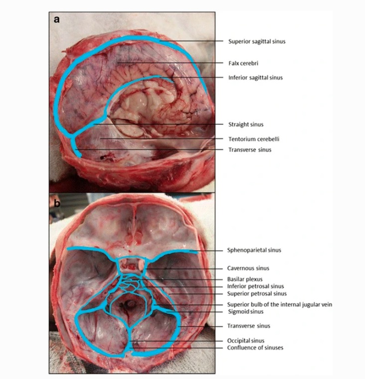
00:01 Let us now move on and look at the meninges of the brain and also the spinal cord. 00:06 Here is a section on the left hand side showing you a section through the skull, skin on the surface, the bone of the skull and then underlying is connective tissue. The periosteum is the capsule that lines the surface of bones and that is continous with the outer layer or the outer meningial layer of the brain, the dura. The dura mater is really almost part of the periosteum. It is the outside covering of the brain. And on the image on the right hand side, this is the section through some neural tissue happens to be the optic tract, but it is a good section to illustrate these meningeal layers. And on the outside it is very thick connective tissue, which is the dura mater. When you have the arachnoid layer, the arachnoid layer has peeled away from the surface of the dura here and that surface of the dura between the arachnoid and the dura is often called the subdural layer, it is an artificial space. Underneath the arachnoid layer is the sub-arachnoid space and on the diagram, you can see that that's a fairly large space indeed. It is full of cerebrospinal fluid that circulates around the brain and keeps the brain protected and buoyant and also flows around the spinal cord. The sub-arachnoid space also houses some of the large vessels before they pass into the deep substance of the brain. And then the most internal covering around brain tissue is the pia mater. It forms almost an epithelial surface on the surface of the brain tissue and penetrates some distance into the neural tissue particularly carrying very small blood vessels. Well lastly let us look at the ventricles of the brain. You know the brain has big spaces in it and these big spaces are occupied by cerebrospinal fluid. Here you see some ventricles, one on the horizontal section through the brain you saw earlier, huge spaces between brain tissue and on the right hand side, you see it labeled in a histological section. The pink stained region that you see above and below the label are neural tissue. But in those ventricle spaces, you can just see some tissue evident. 02:49 That is called the choroid plexus. The choroid plexus is an extension of the pia wrapping around groups of little blood capillaries. And that choroid plexus produces the cerebrospinal fluid and I said it circulates in the sub-arachnoid space, through all these ventricles, along the spinal cord, and finally it returns to the vascular system to the venous system through structures called arachnoid granulations. Well under high power, on the right hand side, you can see these choroid plexuses within the ventricle. And again that remind you they are just really extensions of the pia wrapping around very very small groups of blood capillaries. 03:41 And those epithelial cells that formed the choroid plexus are responsible for making the cerebrospinal fluid. The very complex mechanism they use and that is one that I won't explain now. 03:55 So let us now summarize what we have just been looking at. Make sure you are aware of the structure of the peripheral nerve, its wrappings, its connective tissue coverings. 04:08 Make sure you appreciate the complexity of both the cerebral cortex and the cerebellar cortex, the different cell layers. Make sure you know the different glial cells, recogonize them and know their functions. And finally appreciate the coverings of the brain that protect the brain and the spinal cord. The dura, the arachnoid and the pia. 04:37 So thank you very much for listening to this lecture. I hope you have enjoyed learning something about the structure of the peripheral nerve and also a little brief overview of the histological structure of the brain.
About the Lecture
The lecture Meninges by Geoffrey Meyer, PhD is from the course Nerve Tissue.
Included Quiz Questions
Which of the following structures is responsible for the production of cerebrospinal fluid?
- Choroid plexus
- Dura matter
- Brain nuclei
- Pia matter
- Periosteal layer
Author of lecture Meninges

Geoffrey Meyer, PhD
Customer reviews
5,0 of 5 stars
| 5 Stars |
|
5 |
| 4 Stars |
|
0 |
| 3 Stars |
|
0 |
| 2 Stars |
|
0 |
| 1 Star |
|
0 |
