Playlist
Show Playlist
Hide Playlist
Macular Degeneration

-
Slides OP Retinal Vessel Occlusion Macular Degeneration.pdf
-
Reference List Pathology.pdf
-
Download Lecture Overview
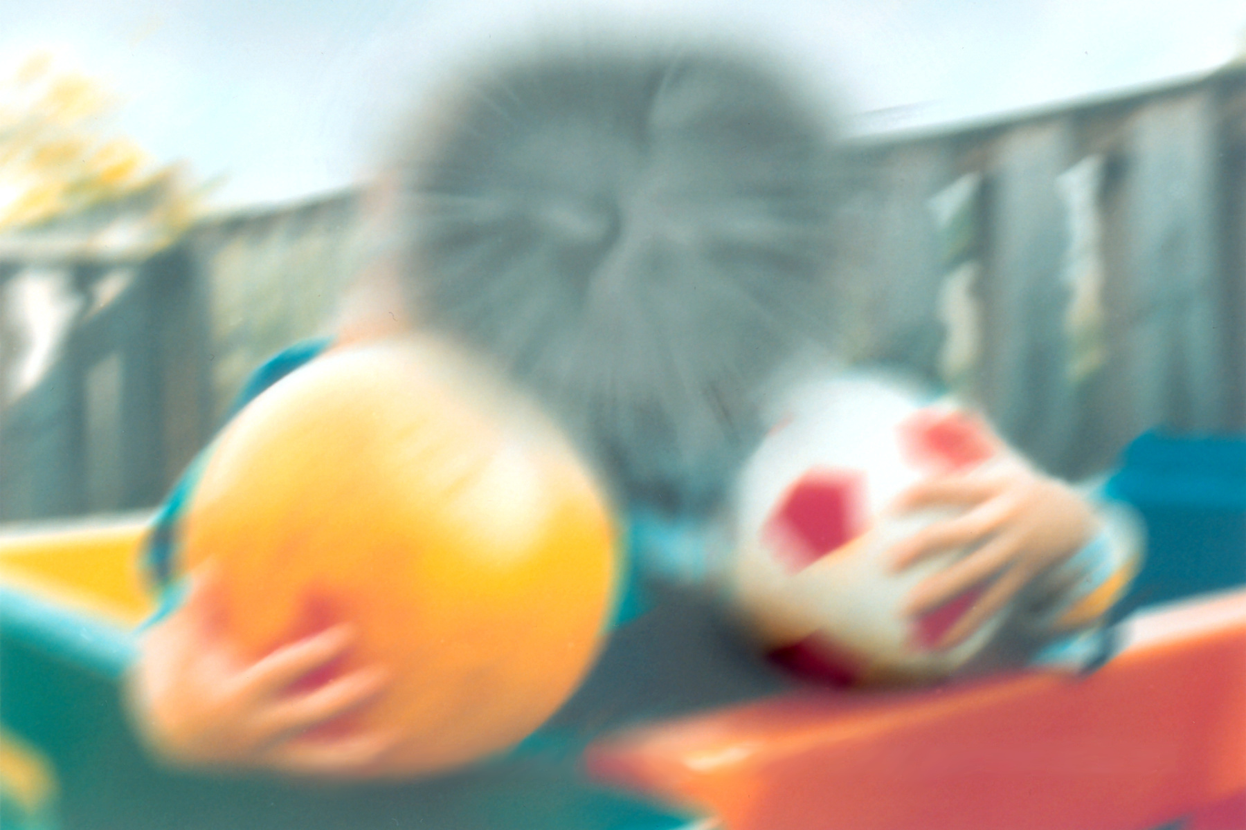
00:00 Let's think about how this is going on in kind of a pathophysiologic way looking at the structures. So, the macula is indicated up above, so that is where we are in the retina and truly that is the highest density of photoreceptors right in that area. Those photoreceptors, those nerves that are going to be collecting the light impulses and then sending it out through the optic nerve are sitting on a layer of retinal pigmented epithelium that sits on a basement membrane that we call, in this case, Bruch's membrane. Because Bruch got their first before basement got their and so he called it Bruch's membrane. Below that is the choroid and below that is the sclera. So that's kind of the general visual 3-dimensional representation of what we're going to be looking at. So, this is what it looks like histologically, I think it's a beautiful structure. Up above those little tiny nuclei that you're seeing are all parts of the rods and cones there in the retina of the patient at the macula. Below that, those single layers of cells that have kind of a dark brown speckled pigment, those are retinal pigmented epithelial cells. And they sit on Bruch's membrane, that's the bright red line that's coming all the way across and labeled Bruch's membrane. And then below that is the choroid. 01:27 Here we've identified it as the choriocapillaris because there is a lot of vasculature and that's going to be providing the necessary nutrition, oxygenation, etc. for the retinal pigmented epithelial cells as well as the overlying rods and cones. So that's what it looks like and that's normal. Okay. Let's make it abnormal in some way. So in AMD, whether it's wet or dry at this point, in AMD we get a deposition of protein and lipids beginning in Bruch's membrane. And again, why we get the deposition we don't entirely understand and why do some patients get and not others. But we begin to see a deposition in that membrane, and that's the membrane that's right below the retinal pigmented epithelial cells that you see identified there. So, we'll begin to get that there. Those deposition, it's proteins and lipids, they are oxidized lipids, there are some complement proteins in there, there are variety of other things. 02:28 They will induce an inflammatory response coming from the choroid. So, the immune system recognizes goes "Hey, those aren't supposed to be there" and they will induce an inflammatory response. Not always a good thing in a small area like this. With inflammation, we get increased vascular permeability and vasodilation in the choroid and we'll get more and more and more accumulation. So, once it get started and once it's underway, it's a little bit of a snowball effect and gradually we can appreciate nodular deposits. And you see them in the bottom panel with a little asterisk, the star associated with them, these are drusen, a German word meaning droplet. And the drusen are the kind of the really recognizable manifestations of these early depositions in Bruch's membrane. Now, they also contain protein and lipid and they're pro inflammatory, they are in many ways helping their manifestation of the inflammation but also continuing to drive in a circular fashion the inflammation. With the inflammatory infiltrates and probably because of free radical damage sometimes associated with the drusen, we get retinal pigmented epithelial cell dysfunction, and they begin to separate themselves from Bruch's membrane. So that's dry AMD in a nutshell. That's dry AMD and that is what is giving us that progressive degenerative loss of visual acuity in that area. 04:12 On the other hand, wet AMD is just building on this, it's only 10% of the population, it's not entirely different in terms of the pathophysiology except in a subset of patients. They will also, as part of the inflammatory response, develop neovascularization. The inflammation will drive vascular endothelial growth factor production which will drive new blood vessels sprouting coming in to the retinal pigmented epithelium from the choroid. Now, those new blood vessels are leaky, they will hemorrhage, they will have much greater amount of exudate, that sort of stuff. So that's the difference between wet and dry AMD, but it all starts in exactly the same way. This is just an example of what that looks like. So, the choroid is below, the retinal pigmented epithelium is above with the retina on top of that. And we have new ingrowth of blood vessels and we have clearly hemorrhage. And so this will give us the wet AMD kind of phenotype. What does it look like on fundoscopic examination? So dry AMD is going to be mainly associated with drusen and the drusen look a little bit like cotton wool spots except they're different. Cotton wool spots, remember our little microinfarcts, the drusen there are these kind of lipid protein depositions that we can see is discrete little things, they're much more broadly distributed in typical cotton wool spots. 05:35 And they're going to be predominantly in the macula because we're getting macular degeneration. On the other hand in wet AMD, there are drusen that are behind there but it's going to be kind of definitely overridden, much more impressive hemorrhage that's associated with that and there will also be neovascularization in earlier stage before the hemorrhage. 05:58 How does it present, signs and symptoms? So macular degeneration is very gradual onset, it is not something that happens overnight. So it kind of sneaks up on you and it will be asymptomatic in most cases. But when it starts becoming symptomatic you find that you will often have bilateral, painless, central visual distortion or loss and so the example there in the bottom panel is very much what it looks like. You can see kind of sort of the faces, but you can't recognize a lot of people. You may even have scotomas, little blind spots that are there. There will be difficulty with night vision because you have lost rods so you have difficulty with low light situations and it takes a long time for you to adapt. So you have to really open up your pupils and collect a lot of light, it takes time to go to get used to be in a low light level. The scotomas will be blind spots in the visual field and again this is going to be right in the middle of your central visual field. You'll have reading difficulty because again the macula is how we read the written word; you cannot make out faces, so the example that you see here the 2 little kids, you can see that there is somebody there holding some balls but you have no idea who. And you can get distortion of those straight lines and that's seen a little bit on the fence behind these kids so metamorphopsia. Okay, and with that we've talked about changes that occur in the vasculature of the eye that can cause blindness, but also a very common entity that we don't completely understand, macular degeneration. Hope you enjoyed it.
About the Lecture
The lecture Macular Degeneration by Richard Mitchell, MD, PhD is from the course Posterior Segment Eye Diseases.
Included Quiz Questions
What are the fatty nodules in age-related macular degeneration (AMD) called?
- Drusen
- Osler's nodes
- Janeway lesions
- Roth spots
- Aschoff bodies
What is the characteristic feature of wet AMD?
- Neovascularization
- Microaneurysm
- Macular detachment
- Ischemic necrosis
- Epithelial hyperplasia
What is seen on the fundoscopic exam in AMD?
- Drusen and hemorrhage
- Drusen and neovascularization
- Drusen and cotton wool spots
- Drusen and hard exudate
- Drusen and cherry red spots
The distortion of straight line that is seen with AMD is called...
- ...metamorphopsia.
- ...metamorphosis.
- ...metaplasia.
- ...metastases.
- ...metachromatic.
Author of lecture Macular Degeneration

Richard Mitchell, MD, PhD
Customer reviews
5,0 of 5 stars
| 5 Stars |
|
1 |
| 4 Stars |
|
0 |
| 3 Stars |
|
0 |
| 2 Stars |
|
0 |
| 1 Star |
|
0 |
Exceptional teacher. Very clear and easy to memorize. Dr Mitchell used very clear didactic tools.
