Playlist
Show Playlist
Hide Playlist
Lumbar Plexus

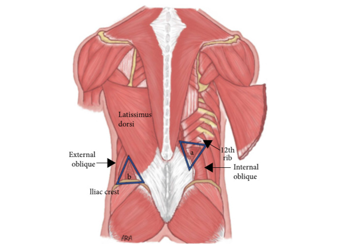
00:00 Remember, we discussed about the brachial plexus in the morning, likewise a few things about the lumbar plexus, lumbar and lumbosacral plexus. 00:12 Who knows about the lumbar, lumbosacral plexus? Tell me anything. What are the nerves, nerve roots? Where do they come? Where do they go? That's sacral. Sorry, it’s L3 down to S2. 00:27 Anything before that, L1? What happens to L1? L1, L2, L3, L4, L5, S1, S2, S3, nerves just coming off this are called the lumbar plexus. 00:51 Nerves coming off this are called the sacral plexus, and coming off both of them is the lumbosacral plexus. 00:58 You don't have to stand up. 01:02 If you remember your anatomy up to the umbilicus, that corresponds to T10. 01:09 Then slightly lower down is T11, T12. 01:13 What is Ll? What are their roots? Can you think of any nerve root of Ll? What about the sensation to the groin? Yes, your ilioinguinal nerve, that's the L1, ilioinguinal, iliohypogastric, iliohypogastric. 01:43 Then you mentioned the genitofemoral nerve, this L1, genitofemoral nerves from L1, L2. 01:58 Is there anything else you can think of? Have you come across a condition called meralgia paresthetica? Yeah. So that also usually comes from L2, L3. 02:12 L2, L3, it runs under the anterior superior iliac spine. 02:17 What's that? Yeah. You get compression, and ascites, pregnancy, or anything compressing on it and cause altered sensation to the lateral aspect of the thigh. 02:27 So, that is lateral cutaneous nerve of the thigh. 02:36 Yes. That's right, usually L1, L2. 02:44 These are the important nerves which are all cutaneous. 02:51 Your genitofemoral nerve has two branches, the genital branch or genitofemoral and the femoral branch. 02:57 The genital branch or genitofemoral is the one which supplies the scrotum or the external genitalia in females. 03:03 The femoral branch is the one which supplies sensation over the anterior aspect of the femoral triangle. 03:11 So that's why when you are testing for dermatome, that's the Ll. 03:16 Now, we'll go to mixed nerves. 03:19 That is L1, L2, L3, sorry L1, L2 supplying. 03:23 Then from L2, L3, L4, those unite and it forms an anterior division and a posterior division. 03:35 Anterior division is the one which forms the obturator nerve. 03:39 Posterior division is the one which forms the femoral nerve. 03:42 So, anterior division is the nerve to the anterior compartment. 03:49 Posterior division is a nerve to the middle compartment. 03:53 So that is L2, L3, L4. What's next? Okay, Hannah. 04:05 Posterior branch. 04:05 Posterior femoral and it branches to obturator. 04:10 So the anterior branches along the middle compartment? That’s right, middle compartment and the posterior one is femoral which is supplying this compartment. 04:16 So, is it medial? Is it posterior? Okay, what else is there? Then anything else? Sciatic. 04:27 Sciatic, sciatic is L4, L5, S1, S2, S3. 04:36 We’ll discuss sciatic in a bit of detail in a minute. 04:39 But for now, just remember this. 04:45 Sciatic, anything else? A couple of other important nerves? You have the superior and the inferior gluteal nerves. 04:55 Superior gluteal nerve comes off 4, 5, S1. 05:01 Inferior gluteal nerve comes off L5, S1, S2. 05:06 You don't have to know the root values. 05:08 But just remember from these plexus, you have the inferior and the superior gluteal nerve supplying all the gluteus muscles. 05:15 The final which will be your S2, S3 for pudendal nerve. 05:24 Pudendal, what does it supply? External urethral sphincter, pelvic diaphragm, levator ani those sort of areas. 05:39 So, S1, S2, S3, S4 keeps your rectum of the floor. 05:45 S2, S3 is always pudendal. That’s your essentially lumbosacral plexus. 05:52 Now, if you go back to the leg, I'm just thinking how to do it quickly without you standing for too long. 06:00 I'll just go through theory a bit more and then you can stand up. 06:03 We've done the anterior compartment, medial compartment, posterior compartment. 06:08 What are the muscles in the posterior compartment? Semimembranosus, semimembranosus, semitendinosus, and biceps femoris. 06:16 So on the lateral side, you have biceps femoris and on the medial side, it’s semimembranosus, semitendinosus Nerve supply: sciatic. 06:25 Now, the biceps femoris, bi, two, biceps femoris has got a long head and a short head, the long head of biceps femoris and the short head of biceps femoris. 06:35 Usually, it is by the sciatic, but occasionally the short head of biceps femoris comes from the tibial nerve. 06:44 Sciatic nerve, I think I won't ask you the sense. 06:49 Sciatic nerve comes down right in the midline of the leg, comes up to the top of the popliteal fossa, and divides into tibial and common peroneal. 07:11 Similar to your radial nerve in the arm which supplies the anterior and posterior aspect, the sciatic nerve supplies the anterior and posterior aspect of the leg. 07:23 One important thing you have to get as a concept is your femoral nerve supplies nothing below the knee. 07:32 Everything below the knee, ankle, foot, it's all from the sciatic. 07:37 That's why it's such an important nerve. 07:39 Femoral nerve’s only job is to supply the hip flexors, so iliacus, psoas, pectineus, then the anterior compartment of the thigh which extends to the knee, that's it. 07:51 Then it's cutaneous, okay? Everything to do with the rest of the knee and the foot, sciatic. 08:04 Popliteal fossa. 08:09 Maybe one more time, we will be able to finish it off, yeah. 08:13 Do you mind? That’s fine. 08:14 Can you turn around please? So very quickly, I don't want him to stand there for too long. 08:19 Very quickly, the popliteal fossa, boundaries, content, it’s similar to the cubital fossa. 08:24 It is quite important. Biceps femoris, semimembranosus, semitendinosus, medial head of gastrocnemius, lateral head of gastrocnemius. 08:33 Just flex, bend your knee a bit. 08:35 So that's the skin. What else is there, skin, subcutaneous fat? That's deeper, but no, on the skin itself. 08:47 Remember, we discussed the cubital fossa. 08:49 I said you have median cubital vein, subcutaneous nerves. 08:53 Anything can you think of here on a skin? I'm sure you can think of-. 08:56 Cutaneous nerves. What cutaneous nerves? I'm sure you can think of-. 08:56 Cutaneous nerves. What cutaneous nerves? Sural nerve, sural nerve, you have the sural nerve. 09:02 Sural nerve comes off the cutaneous and lies in the midline. 09:05 And what vein? You know this. 09:08 Not popliteal vein, short saphenous vein. 09:13 Yeah, you have a short saphenous vein starting from the lateral arch of the foot coming this way along with the sural nerve and emptying the popliteal vein here. 09:25 So they are all on the surface of the skin, on the skin and the popliteal fossa. 09:32 Now, if you reflect the surface, what is the first thing you come across? Imagine, you reflect the skin. 09:40 What is the first anatomical structure, nerve, artery, or vein? Nerve, no, you come across the tibial nerve first. 09:49 Then you have the common peroneal nerve which would have just sort of given off there, common peroneal nerve, also called the common fibular nerve. 09:59 Now it’s called the common fibular nerve or the common peroneal nerve. 10:03 Below that you come across the popliteal vein, and below that, popliteal artery. 10:09 So the deepest structure in there. 10:10 There are two nerves there. 10:11 There’s the tibial nerve and the common peroneal nerve. 10:14 So superficially, you’ve got sural nerve but then you’ve got two further deep. 10:18 That’s right. On that note, I’ll just clarify this. 10:22 The sciatic nerve comes down here, that way. 10:26 Now, about 5, 6 centimeters above the joint line, that's when it divides into two. 10:31 So sometimes you might not see the common peroneal nerve lying there because it's already gone off. 10:35 So you might see only the tibial nerve. 10:37 Tibial nerve, you'll definitely see because it is the midline. 10:40 Common peroneal, you might not see because it might have gone off. 10:43 In your exam, if they ask about the popliteal fossa, they are expecting you to know the tibial nerve, the most superficial. 10:50 Deeper to that is the popliteal vein. Deepest is popliteal artery. 10:55 What’s the floor of the popliteal fossa? Remember, you have a muscle here, brachialis. 11:06 Can you think of a muscle there? Popliteus, popliteus, okay? So simple, popliteus. 11:11 Then you have the capsule of the knee joint. That's it. 11:17 Just turn around for a minute please, a little bit. 11:19 So this is your common peroneal nerve, winding around the neck of the fibula or the common fibular nerve. 11:26 This is the one which divides into superficial peroneal and deep peroneal. 11:31 Superficial peroneal supplies the lateral compartment of the leg. 11:38 Deep peroneal supplies the anterior compartment of the leg. 11:42 These are the two nerves. 11:44 Your tibial nerve at the back supplies all the muscles in the posterior compartment. 11:52 Do you mind just turning around one more time? So, that's your deep peroneal nerve. 12:01 Just think about what you think the function of the deep peroneal nerve is then. 12:04 So, it is to dorsiflex the foot. 12:07 What’s the other action of the deep peroneal? Evert. Evert the foot, correct. 12:13 Similar to what we discussed, we spent a lot of time in the upper limb. 12:18 Likewise, this part of anatomy is very important. It comes up. 12:21 Inversion of the foot, eversion of the foot, at what joint does it happen, which nerve does all that. 12:26 So you need to just concentrate on this bit. 12:28 So your deep peroneal nerve supplies the anterior compartment of the leg. 12:33 What are the muscles in the anterior compartment of the leg here? Tibialis anterior, tibialis anterior, extensor hallucis longus, extensor hallucis longus, peroneus tertius, peroneus tertius One more, extensor digitorum longus. 12:47 So, if his deep peroneal nerve is injured, none of them will work. 12:58 Patient is going to have a foot drop because the dorsiflexors are not working. 13:05 But the plantar flexors are working because that is supplied through the back. 13:09 Will the patient be able to evert the foot? Evert? Evert. Why not? You can invert the foot, but what about eversion? You're right. Common peroneal does both. 13:33 But the deep peroneal does only inversion. 13:36 So if you have a common peroneal nerve injury here, you're right. 13:40 No dorsiflexion, inversion is lost, eversion is lost. 13:46 But if you have a deep peroneal nerve injury there, you have dorsiflexion is lost, inversion is lost, but the eversion is preserved because the superficial peroneal nerve will help in the eversion and it's vice versa. 14:00 So if you have only that nerve injured, only the eversion is lost. 14:05 So if you get a clinical scenario whereby they say everything is okay except that patient is unable to evert the foot, then it is very specific for superficial peroneal or superficial fibular nerve. 14:18 So, the two muscles in the lateral compartment of the leg are peroneus longus, peroneus brevis. 14:26 For the last time, if you would turn around.
About the Lecture
The lecture Lumbar Plexus by Stuart Enoch, PhD is from the course Upper Part of the Body Anatomy.
Author of lecture Lumbar Plexus

Stuart Enoch, PhD
Customer reviews
5,0 of 5 stars
| 5 Stars |
|
5 |
| 4 Stars |
|
0 |
| 3 Stars |
|
0 |
| 2 Stars |
|
0 |
| 1 Star |
|
0 |
