Playlist
Show Playlist
Hide Playlist
Liver: Summary

-
Slides Digestive system liver gallbladder and pancreas.pdf
-
Reference List Histology.pdf
-
Download Lecture Overview
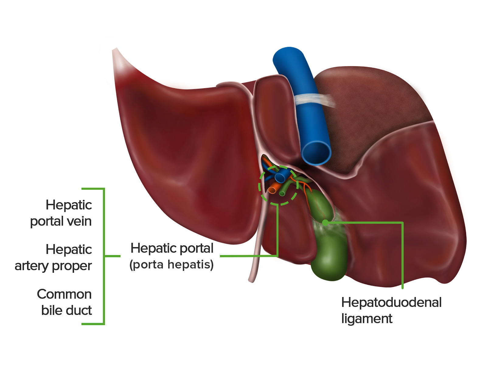
00:01 So in a review, this diagram just summarizes the structure of the hepatocyte. 00:07 Be very careful and very sure that you can identify the different borders, the bile canaliculi and the space of Disse, and identify the enormous factory inside the hepatocyte, and the details of the sinusoids and the microvillus projections of the hepatocyte into the space of Disse and the fenestrated endothelium letting fluid leaking to that space. And also, the macrophage, the Kupffer cell that sits in the endothelial lining an acts as a phagocyte, and also it has other functions. So this is a good summary slide of the function of the hepatocyte and of its relationship to adjacent hepatocytes and also the sinusoids. And I show this slide just to try and summarize the blood flow, first of all, to the liver, and then the flow of the bile from the liver into the gall bladder. If you focus on the right-hand component, the right-hand side of the diagram where there's a very small lobe of the liver, and look at the labels 1 and 2, it does illustrate one part of the blood supply to the liver that's important. Portal vein carries blood from the intestine. 01:36 It goes into the liver, as you see here, and branches into a number of interlobar and interlobular veins. And they eventually find their way to the corners of these portal triads where they branches at the portal vein. 01:54 And that blood pours towards the central vein, the contents get processed by the hepatocytes, the hepatocytes return some components to the blood, and the blood, when it drains to that central vein, then passes out of the liver through the inferior vena cava back to the heart, and then to circulate around the body. On the left-hand side, on the larger lobe, it illustrates two things, the arterial supply to the liver and the duct or the hepatic duct and the bile duct drainage from the liver. 02:32 First of all, look at the hepatic artery. It supplies blood, oxygenated blood to the liver, and then branches to form that branch to the hepatic artery that is going to join the venous blood coming from the portal vein, and circulate into that central vein past the hepatocytes and supply them with oxygen. Now, focus on the green structures. They are the bile ducts, the bile canaliculi carried by or made by the hepatocyte back to the edges of the portal triads, and then they eventually find their way into the hepatic ducts. And a number of ducts follow and are named according to their locations. And finally, the bile goes and gets stored in the gall bladder, where it's stored, concentrated, and then released through the common bile duct into the duodenum. Notice on this slide also that there is a pancreatic duct, also going into the common bile duct, also enabling the pancreas to deliver its exocrine secretions, digestive enzymes through that duct into the duodenum.
About the Lecture
The lecture Liver: Summary by Geoffrey Meyer, PhD is from the course Gastrointestinal Histology.
Included Quiz Questions
Which of the following vessels supplies fully-oxygenated blood to the liver?
- Hepatic artery
- Mesangial artery
- Central vein
- Portal vein
- Hepatic vein
Which of the following cells in the liver are also known as stellate macrophages?
- Kupffer cells
- Histiocytes
- Dust cell
- Langerhans cell
- Microglia
Author of lecture Liver: Summary

Geoffrey Meyer, PhD
Customer reviews
5,0 of 5 stars
| 5 Stars |
|
5 |
| 4 Stars |
|
0 |
| 3 Stars |
|
0 |
| 2 Stars |
|
0 |
| 1 Star |
|
0 |
