Playlist
Show Playlist
Hide Playlist
Vision: Lens, Pupil and Humors
-
Slides 02 Vision NervousSystem.pdf
-
Download Lecture Overview
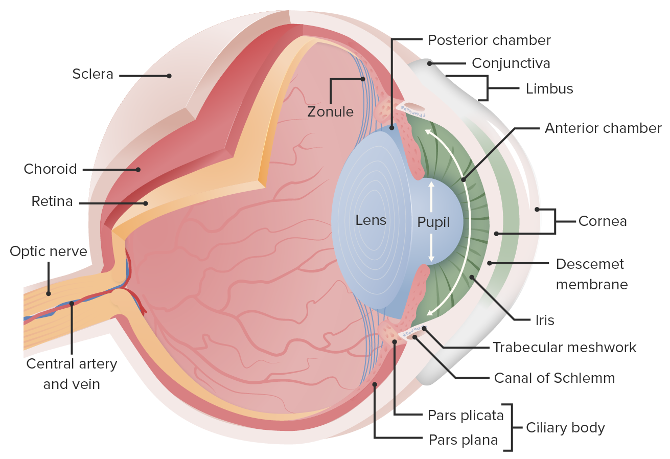
00:00 It’s probably best for all of us to think of the eye like a camera when we first start. 00:07 This gives us some working knowledge of what things we’re gonna be able to change whether eye to get the best image possible to the brain. 00:16 And it is very important to get that best image possible, both in terms if you were trying to forge for food or runaway from a predator or even have a good social interaction with a neighbor or colleague. 00:28 So if you think of the eye being like a camera, what are the couple components that the camera that we can now relate to the eye and the eye structure? One of the big one is the aperture. 00:39 Aperture is the whole in which the light travels through. 00:43 And you can alter that to get more light into a camera or to get more light into the eye. 00:51 The other interesting or the important thing is you can adjust the focus of the lens so that you can get an optimal clarity of the image. 01:01 Those are two important items and your eye does the very same thing, by altering the pupil with is the same as the aperture and altering the lens is the same as adjusting the focus. 01:16 In addition, to the lens, you also have some sort of film or way to record the image. 01:24 So in the olden days when we use photography film to put the image on, that is, think of that like the retina, its collecting the information that’s being sent through a cornea, the various humors, into the lens and then back to back to the eye. 01:42 That photography or photographic film is the same as photoreceptors. 01:53 So, going through these humors and lenses. 01:56 We always need to go through what is the path of the light has travelling to get back to the retina. 02:03 The aqueous humor is that first humor that we have for fluid that will eventually be drained into a canal. That will then be collected up by the lymphatic system. 02:16 And remember we talked about before you need to have that flow from the posterior chamber through the pupil into the anterior chamber before its drained. 02:27 Finally, to think about is that various humors will reflect the light. 02:33 What is light refraction mean? It means that the light is bent slightly. 02:40 So for every object you travel through, you’ll need to account for how much that light is being bent. 02:48 So air versus the cornea, versus the aqueous humor, versus the lens, versus the vitreous humor. 02:56 All will just the light bend. 03:00 that needs to be accounted for when you were showing an image to the back of the retina. 03:08 What kind of process that occurs? The less light bending the better. 03:15 And that is why you don’t have structures that are in place that allow the light to travel through that absorb that light. 03:22 Because you don’t want any, what we call chromophores which absorbs the light because you want that light to pass through with the least bending as possible. 03:33 Now the lens itself also refracts light but it can accommodate. 03:38 What we mean by accommodate is change width and how much it is stretch so that you can adjust the focal length to the back of the retina. 03:50 This allows you to see both near things, readjust see far things, readjust see near things again. 03:59 It is how you’re moving your focus in and out because you can’t focus on both near and far things at the same time. 04:08 Just like with your camera, you have to adjust the lens to look at near things. 04:13 You have to adjust the lens to look at far things. 04:17 How that adjustment looks? Is all in terms of muscle contractions on the lens itself. 04:24 So you have ciliary muscles that will either contract or relax. 04:29 If they are contracting, they are relaxing the lens. 04:34 If they are relaxed, the lens tenses up. 04:39 That may seemed counterintuitive at first. 04:42 But remember, you have these little fibers called zonule fibers. 04:46 and these are pulling on the lens at all times. 04:50 So actually, by contracting the ciliary muscles you're relieving that tension being pulled on with these zonule fibers. 04:59 How do you adjust pupil size? Pupil size is the aperture how much light you’re getting through the pupil is gonna be a very important for how bright your image is. 05:11 If you have high light levels, the sphincter muscles are going to constrict to get smaller and that decreases the pupil diameter and how much light can move in. 05:26 If light levels are too low, the opposite happens. 05:29 You have muscles are called radial muscles. Those will constrict which dilate the pupil. 05:38 Why is this sympathetic parasympathetic component important? Well sympathetic nervous system response is to cause a pupil dilation and a parasympathetic nerve response is to cause the pupils constriction. 05:52 These are always balance in each other. 05:56 So in fact, they’re always have one, both of these play at the same time. 06:02 You can adjust it one direction or another if you do something like a pupillary light test. 06:07 You take a small flash light and just move it towards the eye. 06:10 You can get the eye to change size in terms of the pupil. 06:15 The physician will often times utilize this principle of having both of these in play at the same time. 06:23 Because you can remove one or the other to get the opposite response. 06:29 So for example, a parasympathetic nervous system response to cause a sphincter muscle constriction is using acetylcholine. 06:38 Therefore, you can give a blocker of acetylcholine like atropine and get dilation of the eye. 06:46 So you can block the parasympathetic to get pupil dilation. 06:50 because these are always going on at the same time. 06:53 These remember are just the aperture or how much light gets into the eye. 06:59 Therefore, if you have your pupils dilated by something like atropine you might need to wear your some sort of sun protection as you go outside because your pupils will be dilated.
About the Lecture
The lecture Vision: Lens, Pupil and Humors by Thad Wilson, PhD is from the course Neurophysiology.
Included Quiz Questions
In which sequence does light enter and course through the eye?
- Object - air - cornea - aqueous humor - lens - vitreous humor - retina
- Retina - vitreous humor - lens - aqueous humor - cornea - air - object
- Object - lens - aqueous humor - vitreous humor - retina
- Object - lens - air - retina
- Air - Object - lens - retina
Which of the following would cause the lens to relax?
- Contraction of ciliary muscles
- Relaxation of ciliary muscles
- Taut zonule fibers
- Viewing an object far away
- Bright light
What acts as an aperture to restrict light entry into the eye?
- Iris
- Cornea
- Retina
- Lens
- Sclera
Customer reviews
4,0 of 5 stars
| 5 Stars |
|
0 |
| 4 Stars |
|
2 |
| 3 Stars |
|
0 |
| 2 Stars |
|
0 |
| 1 Star |
|
0 |
le falta un poco más de información, pero como algo general está bien.
very good diagrams, would have liked to see an even deeper dive into ciliary muscle in relation on zonule fibers and how they are inverse to each other,


