Playlist
Show Playlist
Hide Playlist
Lens
-
Slides 16 Human Organ Systems Meyer.pdf
-
Reference List Histology.pdf
-
Download Lecture Overview
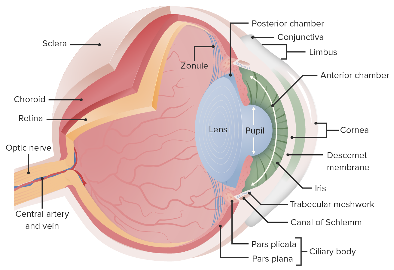
00:01 So let us look at the lens now in more detail. On the left-hand diagram, it is a large picture. You can see the zonular fibre extending from the ciliary epithelium of the ciliary processes and then you see the structure of the lens. It has got a capsule all the way around it, a clear transparent capsule and then underneath that capsule is an anterior epithelium, the lens epithelium. It is only on the anterior surface. The posterior surface does not have that epithelium and those anterior lens epithelial cells produce the lens fibres and those lens fibres if you look on the right-hand picture there you see at the equator that epithelium produces the lens fibres and then those fibres move towards the centre of the lens and their nuclei as they move towards the centre start to disappear and finally end up as what we call ghost nuclei. They are just little spaces that are finally filled up. Now these lens fibres to be anything up to 8 to 10 millimeters in length. 01:18 They are about 8 t 10 microns in width and they are packed all together and as they move towards the nucleus after being constantly produced at the equator, they tend to fuse together and its very hard to separate and will see the different fibres lined up. And as they move towards the centre, they also accumulate the protein crystallines. These lens fibres are produced regularly. They consist of type IV collagen and is very elastic and that elasticity changes with age. 02:00 It gets reduced as we get older. When you are in the fourth decade of your life suddenly the elasticity changes so the lens can no longer accommodate, change its shape as well as what it did in the younger person. That is called presbyopia and therefore, the person normally has to wear glasses to accommodate for that, to assist the lens to focus the light rays on the retina. You can see here again the zonular fibres extending from the ciliary processes to the lens and this summarizes what happens when the ciliary fibres are stretched or put tension on or they relax caused by the ciliary muscle. On the left-hand side when the ciliary muscle contracts, the ciliary body squeezes up and moves closer towards the lens. 02:59 So the zonular fibres become more relaxed and so the lens will flat out. It will become wider. Conversely when the ciliary muscle is relaxed, then the ciliary body moves away from the lens and the fibres become stretched or tensed and, therefore, the lens get thinner change its shape. And this is how the lens, therefore, can focus light from distant objects or closer objects regularly onto the surface of the retina, the process of accommodation.
About the Lecture
The lecture Lens by Geoffrey Meyer, PhD is from the course Sensory Histology.
Included Quiz Questions
Which of the following regarding the microanatomy of the lens is MOST ACCURATE?
- The lens epithelium is located in the anterior portion of the lens between the lens capsule and the lens fibers.
- The lens epithelium completely surrounds the lens.
- The lens capsule is located in the anterior portion of the lens between the lens epithelium and the lens fibers.
- The lens capsule is located in the posterior portion of the lens between the lens epithelium and the lens fibers.
- The lens epithelium is located in the posterior portion of the lens between the lens capsule and the lens fibers.
Presbyopia is most commonly caused by which of the following?
- Aging
- Glaucoma
- Excessive light
- Trauma
- Infection
Customer reviews
5,0 of 5 stars
| 5 Stars |
|
5 |
| 4 Stars |
|
0 |
| 3 Stars |
|
0 |
| 2 Stars |
|
0 |
| 1 Star |
|
0 |


