Playlist
Show Playlist
Hide Playlist
Leishmania – Protozoa (Visceral Infection)

-
Slides 12 Leishmania MicrobiologyAdvanced.pdf
-
Download Lecture Overview
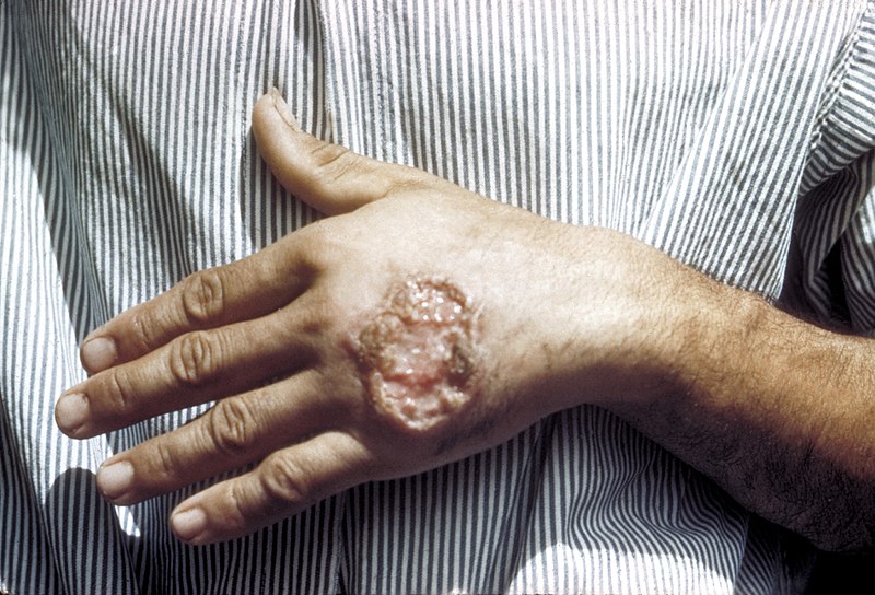
00:01 Hello, and welcome to Leishmania. 00:04 This is one of a series of lectures where we look in depth-at individual human parasites. 00:10 After you hear this lecture, you'll understand the life cycle of Leishmania and how it's transmitted to humans. 00:18 You'll be familiar with the pathogenesis of the different forms of leishmaniasis, and you'll understand how to prevent and treat leishmaniasis. 00:31 We've been looking at a series of protozoan parasites? and just to remind you, these are single-celled eukaryotes of medical importance. 00:44 We looked at plasmodium, toxoplasma, and now, let's take a look at the leishmania. 00:51 Here's a photograph of the blood form of leishmania. These are flagellated protozoan parasites and leishmania come in a variety of species which are medically important. 01:04 These species have different disease outcomes and have different global distributions. 01:11 On the left, you have the distribution of a disease we call cutaneous leishmaniasis. 01:19 This is a disease as you will see that mainly affects the skin. 01:24 And on the right is visceral leishmaniasis where the parasite causes disease in your internal organs. 01:33 And you can see, by just the glance at these two maps, that the distributions are overlapping but not identical. 01:41 If we look at cutaneous first on the left, the darker countries have more cases. 01:46 So, you can see Central and South America have a number of cases. 01:50 Africa, the northwestern part of the country, and Egypt, Central Asia, and that's cutaneous. 01:59 Now, on the right, if you look at visceral leishmaniasis, you see Central and South America as well but now, a different distribution on the eastern part of Africa and particularly high levels of cases in India. 02:14 And these are reflection of, in part, the animals that are infected and the vectors that transmit them. 02:21 Now, one of the species, Leishmania donovani, causes visceral leishmaniasis. 02:28 It's a very serious disease. 02:30 There are about 300,000 cases a year and as you see on the map on the right, the lower right,?many of them in South America, Eastern Africa, and India. 02:42 300,000 cases per year and 20,000 deaths. That's Leishmania donovani. 02:49 Two strains, Leishmania tropica and Leishmania mexicana, cause cutaneous leishmaniasis. 02:58 This is, again, a lesion in the skin. A million cases in the past five years or so. 03:04 That's the graph on the top and this is not a life-threatening disease but the lesions can be disfiguring. 03:11 If they occur on the face, you will have a lesion for the rest of your life and it will be seen, of course. 03:17 And the fourth strain, Leishmania braziliensis, causes muco-cutaneous leishmaniasis. 03:26 These are lesions on the skin but they also tend to travel to muco-cutaneous membranes?as we'll see in a moment. 03:35 Leishmania are obligate intracellular parasites. They need to get inside of cells in order to reproduce. 03:42 Although, of course, there are extracellular faces in which the parasites can move about the body. 03:49 All of these leishmania infections are transmitted by the bite of a phlebotomous sandfly. 03:57 Now, you may know that the word 'phlebotomous' means blood-drawing? and that's because these flies like to drink your blood and have it as a meal? and in so doing, they transmit leishmaniasis. Leishmaniasis are zoonotic infections. 04:15 They are principally infections of animals of various kinds. 04:20 The human becomes infected when the vector transmit the parasite from an animal to a human. 04:26 Now, some transmission does occur from human to human in areas where the parasite is endemic? but for the most part, it's considered as zoonotic infection. So, here's the life cycle of Leishmania tropica. 04:43 Now, remember, this parasite can be acquired largely from other animals. 04:48 The horse, dog, animal, but it can also be passed from human to human? and again, the phlebotomous sandflies pick up the parasites, deliver them to humans,? and then they are passed around to other animals as well. 05:02 Wherever the sandfly bites, that's where the lesion occurs. If it's on your face, you're left with a scarring lesion. 05:09 So first, here is a sandfly picking up the amastigote form of the leishmania, which it can do from many different mammals. 05:19 On the top right, non-human animals and human animals as well. 05:24 It takes a blood meal, it pulls up the parasites into its gut, of course,? and there, it will undergo a number of transformations and make its way back to the salivary gland? so that it can be delivered through the saliva to another host. 05:41 The sandfly acquires the amastigote form that's shown on the lower right. 05:46 That form goes to the gut tract and then transform into flagellated promastigotes. 05:54 These are freely swimming forms of the parasite. 05:58 They replicate in the gut tract, make their way to the salivary gland? which you could see in the middle circle there, and that's how they get delivered to a new host. 06:08 Of course, if they remained in the gut tract, they wouldn't get delivered to a new host? because the contents of the gut are not injected when a biting animal or a biting fly bites a new host. 06:20 And so then, once the parasites have developed and moved to the salivary gland? when the sandfly bites a new host they can be injected into another animal. 06:30 And as I said, very important observation, in endemic areas,? that means areas where there are lots and lots of leishmania infections of humans,? it is possible that the sandfly could bite an infected human, pick up leishmania parasites, and deliver it to another human or perhaps, even another animal that it's biting. 06:50 This has to be an area of very high density infections because as you can imagine, if there are just a few human infections,? it's not likely that a sandfly bite is gonna pick up the parasite from them. 07:01 More likely picking it up from animals which are more heavily infected. 07:06 Once the sandfly injects the parasite, the flagellated form into the skin,? it leads to tissue destruction and the formation of a lesion at the site of the bite,? which you can see on the hand shown here. 07:23 And this is, in part, due to the fact that the flagellated forms are taken up by macrophages,? they replicate in the macrophage, the macrophage is destroyed,? it releases a variety of chemicals that can then destroy the surrounding tissues. 07:37 So, this can be a lesion of an intra-cell diameter. 07:41 On the hands, it's of no consequence but of course, it often happens on the face. 07:45 You can be bitten there and these are typically not treated and you have a scar for the rest of your life unfortunately. 07:54 So, that's cutaneous leishmaniasis. 07:57 Now, Leishmania braziliensis, causes what's called muco-cutaneous leishmaniasis? and the life cycle is for the most part, very similar. A mosquito has picked up the amastigote form, the amastigote transforms?into a flagellated form in the gut of the mosquito,? it then moves to the salivary gland, the mosquito I'm sorry, I'm saying mosquito but I mean sandfly, of course. 08:27 The sandfly then bites another host, a lesion forms at the side of the bite through the same mechanisms that we've discussed. 08:36 But for L. braziliensis, the infective macrophages containing amastigotes,? then leave the site of the initial bite and move to muco-cutaneous membranes? either in the oral pharynx or in the genital tract. And those two are diagram at the bottom of the picture. 08:58 There, additional tissue destruction occurs forming the muco-cutaneous lesion. 09:04 And these can be quite horrific. 09:06 Here's a photograph of a gentleman whose nose has been eroded away? by this kind of replication and tissue destruction within macrophages. 09:16 That's a muco-cutaneous lesion. This can occur in the mouth or in the genital areas. 09:22 So, you can imagine, if you simply search for this condition online, you will find horrific photos of people who are - who have amazingly degenerated muco-cutaneous lesions like this one. 09:35 So, that's Leishmania braziliensis. 09:38 Then, we have visceral leishmaniasis which is the more serious disease in terms of mortality?and this is caused by Leishmania donovani. 09:48 And again, the life cycle is very similar. 09:52 We have sandflies delivering flagellated forms to the tissues by a bite, again,?but these enter macrophages as before. 10:04 You don't have a lesion forming at the bite site? but then, these macrophages go to other tissues such as the liver and other internal tissues. 10:13 They deliver the parasites to those organs where they replicate, induce cell death, and damage the organs. 10:21 That's why we call it visceral leishmaniasis. 10:25 And once again, the sandflies can pick up the flagellated forms or the amastigote forms from the infected host whether it be human or an animal and start the cycle all over again. 10:37 So, this is visceral leishmaniasis and again, this is the one associated with substantial death. 10:44 And the manifestation of visceral leishmaniasis, the disease is called Kala-azar,? which means black fever because the skin of the victims often turns a dark color at the height of infection. 10:59 It has a three to six-month incubation period after the time that the sandfly delivers the parasite by a bite. 11:08 It takes three to six months for the symptoms of Kala-azar to become evident. 11:13 And after the black skin or the black appearance of the skin that gives the disease its name,? it's associated also with high fever and splenomegaly, swollen spleen,?sometimes swollen liver, and on this individual who has Kala-azar, you can see the spleen and liver have been outlined. 11:33 And you can see his belly is very distended as a consequence of this damage?caused by replication of the parasite. 11:41 Now, this infections also associated with anemia. 11:46 You can also have congenital Kala-azar if you're infected while pregnant. 11:51 The parasites, of course, have the ability to go all over in the circulation. 11:55 They can certainly enter the fetus and cause substantial damage there as well. 12:00 So, this is something you absolutely want to avoid but in areas where the vector is present and the parasite is present very difficult to avoid without good vector control. 12:11 After this entire infection is over, there's an event called post-Kala-azar dermal Leishmaniasis. 12:20 About 20% of the infections proceed to this point and these are associated with a most unusual presentation. 12:29 Here, we show a gentleman's face which is covered with these puffy swollen lesions and this can occur all over the body.
About the Lecture
The lecture Leishmania – Protozoa (Visceral Infection) by Vincent Racaniello, PhD is from the course Parasites.
Included Quiz Questions
Leishmania belongs to which variety of protozoan parasites?
- Flagellates
- Ciliates
- Sporozoans
- Sarcodines
- Non-ciliates
Which of the following is true about Leishmanias?
- They are single-celled eukaryotes.
- They are multi-celled prokaryotes.
- They are single-celled fungi.
- They are single-celled prokaryotes.
- They are multi-celled parasites.
Leishmania donovani mainly affects which of the following body parts?
- Liver
- Epidermis
- Subcutaneous tissue
- Salivary gland
- Lacrimal glands
Which strain of Leishmania causes mucocutaneous leishmaniasis?
- Leishmania braziliensis
- Leishmania mexicana
- Leishmania tropica
- Leishmania donovani
- Leishmania aethiopica
Which organism acts as a vector for Leishmania?
- Phlebotomus sand fly
- Male Anopheles mosquito
- Sporothrix schenckii
- Ixodes scapularis
- Mansonia mosquito
Which form of Leishmania is taken up by the Phlebotomus sand fly when it bites a host?
- Amastigote
- Promastigote
- Protomastigote
- Schizont
- Parasitophorous vacuole
In which part of the sand fly does Leishmania replicate?
- Gut tract
- Thorax
- Proboscis
- Salivary glands
- Palpi
What causes the destruction of tissue and the formation of a lesion at the site of a bite from a sand fly infected with Leishmania?
- Release of chemicals from macrophages that are destroyed by Leishmania
- Proteases contained in sand fly saliva
- Low pH of sand fly saliva
- Anticoagulants contained in sand fly saliva
- Release of antihistamine from mast cells
After replication, the flagellated form of Leishmania tropica travels to which part of the sand fly?
- Salivary gland
- Thorax
- Proboscis
- GI tract
- Palpi
After engulfing the flagellated form of Leishmania braziliensis, macrophages migrate to which membrane?
- Mucocutaneous membrane
- Basement membrane
- Pleural membrane
- Epidermis
- Hypodermis
After engulfing the flagellated form of Leishmania donovani, macrophages migrate to which part of the body?
- Internal organs
- Skin
- Mucous membrane
- Salivary glands
- Lacrimal glands
Kala-azar is associated with which of the following strains of Leishmania?
- Leishmania donovani
- Leishmania mexicana
- Leishmania tropica
- Leishmania braziliensis
- Leishmania aethiopica
There are how many cases of visceral leishmaniasis per year?
- 300,000
- 200,000
- 100,000
- 400,000
- 75,000
What are the most prominent features of kala-azar?
- High fever and splenomegaly
- Black saliva
- Jaundice
- Renal failure
- Cerebral edema
Author of lecture Leishmania – Protozoa (Visceral Infection)

Vincent Racaniello, PhD
Customer reviews
5,0 of 5 stars
| 5 Stars |
|
1 |
| 4 Stars |
|
0 |
| 3 Stars |
|
0 |
| 2 Stars |
|
0 |
| 1 Star |
|
0 |
much wanted detail. thank you so much, perfect and informative.
