Playlist
Show Playlist
Hide Playlist
Bowel Obstruction and Ileus: Large Bowel Obstruction & Ogilvie Syndrome

-
Slides Ileus and Obstruction.pdf
-
Download Lecture Overview
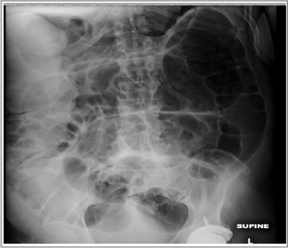
00:00 So let's move on to large bowel obstruction. 00:04 This is usually caused by a mechanical obstruction of the large bowel and the cecum is usually, the part of the bowel that dilates the most. 00:12 The small bowel is often not dilated unless the obstruction is so severe that it makes the ileocecal valve incompetent. 00:19 In a large bowel obstruction, you don’t want to administer oral barium because it could become impacted within the bowel as water is absorbed from it. 00:29 So you wanna start off by doing a plain film and if you suspect a large bowel obstruction then don't administer the oral contrast. 00:36 So let's move on to volvulus. 00:40 A volvulus is essentially a closed loop obstruction involving the large bowel. 00:44 It's most commonly seen in the cecal area and then it's followed by the sigmoid area. 00:50 Sigmoid volvulus tends to produce a characterized "coffee bean" shape appearance on radiographs. 00:55 With the cecal volvulus, you'll actually see a very dilated cecum that often rotates to the left upper abdomen. 01:03 On this film, you can see what appears to be a very dilated cecum. 01:07 It remains within the pelvis in this patient. 01:10 The small bowel will become dilated with volvulus and as with the small bowel volvulus there's a very high risk of strangulation. 01:18 So let's take a look at this film, you can see the green line, showing you a very dilated loop of large bowel, so what is this represent? This is actually an example of a Sigmoid Volvulus and you can see, this is the characteristic coffee bean shape that you would expect. 01:37 This is a surgical specimen demonstrating the same thing, a sigmoid volvulus. 01:43 So a colonoscopy is used to reduce the volvulus most often. 01:47 Occasionally, you can use a contract enema, however you have to be careful because when you reduce the volvulus there is a high risk of perforation. 01:55 So what are other secondary science of volvulus. 02:00 You can see the whirl sign which is swirling of the mesentery and that could be seen with both large volvulus and small bowel closed loop obstruction. 02:09 You can also see what's called the beak sign which is tapering the colon to the point of obstruction. 02:13 So let's take a look at these images, this is an axial CT image in a patient that has a closed loop obstruction and here you can see what's called the beak sign. 02:24 So you can see tapering in to the colon, producing the appearance of the beak. 02:28 And then on the coronal image, keep your eye on this area right here as I scroll through. 02:33 You can see what looks like a swirling of this mesentery and this is another secondary sign of volvulus. 02:40 So Ogilvie Syndrome is actually massive dilatation of the colon without mechanical obstruction. 02:49 So as you recall, normal large bowel obstruction is often due to a mechanical obstruction. 02:54 This is one of the causes of a non-mechanical obstruction than can cause dilatation of the colon. 02:59 Usually this is due to anticholinergics which result in loss of peristalsis, trauma especially retroperitoneal. 03:06 Serious infection and cardiac disease. 03:09 So let’s go back to this case that we saw at the very beginning. 03:12 This is our 25 year old female that presents with right lower abdominal pain. 03:16 What do you see on this film? So we see multiple loops of large bowel that contain air and that’s normal. 03:30 But how about right here? What is that represent? So this is a prominent loop of air-filled small bowel that seen in the left upper abdomen and this is consistent with the focal ileus. 03:50 As you can see here, it has that stacked coin appearance that we talked about. 03:54 So what's the next step? How do you figure out what's going on? Why does this patient have a focal ileus? So you want to obtain of CT of the abdomen and pelvis with contrast to take a look for any kind of causes. 04:07 Remember there are multiple different causes of the small bowel focal ileus. 04:12 So let's take a look at this axial contrast enhanced CT scan. 04:17 And if you look right here, you actually have a very thick walled appendix with periappendiceal fat stranding. 04:28 Let's take a closer look. So this portion right here is all appendix. 04:32 Normally appendix should have a little bit air or a little bit of contrast within it but you actually don't see the lumen at all because the wall is so thick and if you look around it, let's take a look at normal mesentery so this area right here is normal mesentery. 04:47 Surrounding the appendix, the mesentery has a more grayish appearance and this is because of surrounding inflammation. 04:55 So this patient has an acute appendicitis that caused her focal ileus. 04:59 So we've gone over a multiple different causes, of large and small bowel obstruction. 05:04 Again this is a very common finding that you wanna look out for when you see a patient coming in with abdominal pain.
About the Lecture
The lecture Bowel Obstruction and Ileus: Large Bowel Obstruction & Ogilvie Syndrome by Hetal Verma, MD is from the course Abdominal Radiology. It contains the following chapters:
- Large Bowel Obstruction
- Ogilvie Syndrome
Included Quiz Questions
Oral barium is not administered with large bowel obstruction because…?
- …it could become impacted as water is absorbed from it.
- …it could cause a bowel hernia.
- …it could cause strangulation.
- …the bowel wall tends to retain the barium for months.
- …it could give a false diagnosis.
Which of the following is a FALSE statement regarding large bowel obstruction?
- Functional obstruction is the most common cause of large bowel obstruction.
- Cecum often dilates the most.
- A volvulus is a closed loop obstruction involving the large bowel.
- In a cecal volvulus, the dilated cecum often rotates to the left upper abdomen.
- There is a high risk of strangulation.
What is Ogilvie syndrome?
- Massive dilatation of the colon without mechanical obstruction
- Massive dilatation of the cecum with a normal colon
- Functional dilatation of the rectum
- Mechanical obstruction of the colon with dilatation
- Absent peristalsis in the ileum leading to dilatation of the colon
Author of lecture Bowel Obstruction and Ileus: Large Bowel Obstruction & Ogilvie Syndrome

Hetal Verma, MD
Customer reviews
5,0 of 5 stars
| 5 Stars |
|
5 |
| 4 Stars |
|
0 |
| 3 Stars |
|
0 |
| 2 Stars |
|
0 |
| 1 Star |
|
0 |
