Playlist
Show Playlist
Hide Playlist
Intussusception: Examination and Diagnosis

-
Slides Intussusception Surgery.pdf
-
Download Lecture Overview
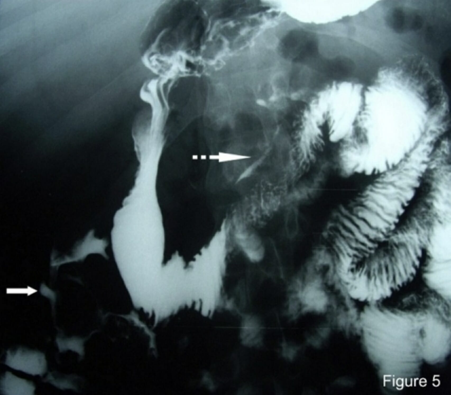
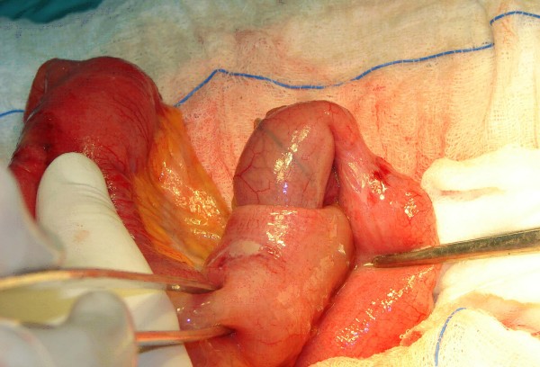
00:01 Physical findings common for intussusception. 00:04 This may be difficult to differentiate from other signs of abdominal pain. 00:10 Patients may vomit. 00:12 Initially, the vomiting is non-bilious. 00:14 This is reflexive. 00:16 Don't be confused. 00:17 This is not a diagnosis of hypertrophic pyloric stenosis, which, as you know, results, in non-bilious emesis and projectile vomiting. 00:26 Later, however, the emesis is usually bilious. 00:29 Now, the emesis is due to a small bowel obstruction. 00:36 The patient may have abdominal pain. 00:38 Particularly with worsening abdominal pain, you should be concerned about ischemia or impending ischemia. 00:46 And sometimes, passage of bloody stools. 00:50 I hope you don't like currant jelly stool because this is what is classically described as bloody stools in intussusception. 00:59 What laboratory findings might you find? On chemistry, due to the vomiting, you may classically find low sodium, low chloride and low bicarb. 01:10 Additionally, particularly with signs of ischemia, you may find a child with an elevated white blood cell count or leukocytosis. 01:19 Let's move on to imaging to help diagnose. 01:22 Ultrasound, remember, is incredibly helpful in the young. 01:26 We try not to expose our babies and infants to radiation as much as possible. 01:31 Again, to remind you, the abdominal wall of a child is usually thin and quite amenable to ultrasound techniques. 01:39 As you can see on this ultrasound image, this is a classic target sign. 01:44 Notice the white circle in the middle of the screen, that's the portion of the intestines that’s invaginated or telescoped into the proximal bowel. 01:54 Also important to notice, the thick black band around the inside circle is edematous proximal bowel. 02:01 Remember our discussion about venous and lymphatic congestion. 02:05 This finding is consistent with potentially ischemic bowel and this patient may need surgery. 02:14 Abdominal x-rays unfortunately are much less reliable. 02:18 However, we’ll shortly discuss why contrast studies may actually be both diagnostic and therapeutic. 02:28 This is a cross-sectional CAT scan of the abdomen and pelvis clearly in an adult. 02:33 I want to point out to you that intussusception can happen in adults as well. 02:37 And it's rare for us to get a CAT scan in children. 02:40 Do you notice the target sign here shown by the white arrow? It looks remarkably similar to the ultrasound, doesn't it?
About the Lecture
The lecture Intussusception: Examination and Diagnosis by Kevin Pei, MD is from the course Special Surgery.
Included Quiz Questions
Which of the following best describes the physical findings of intussusception?
- Non-bilious vomiting later followed by bilious vomiting
- Tenderness of the abdomen at McBurney's point
- Vomiting and diarrhea
- Bilious vomiting later followed by non-bilious vomiting
- Erythema of the abdominal wall
What type of stool quality is associated with intussusception?
- Currant jelly stool
- Mucus in stool
- Green stool
- Clay-colored stool
- Persistent black stool
Author of lecture Intussusception: Examination and Diagnosis

Kevin Pei, MD
Customer reviews
5,0 of 5 stars
| 5 Stars |
|
5 |
| 4 Stars |
|
0 |
| 3 Stars |
|
0 |
| 2 Stars |
|
0 |
| 1 Star |
|
0 |
