Playlist
Show Playlist
Hide Playlist
Introduction – Gastrointestinal Motility
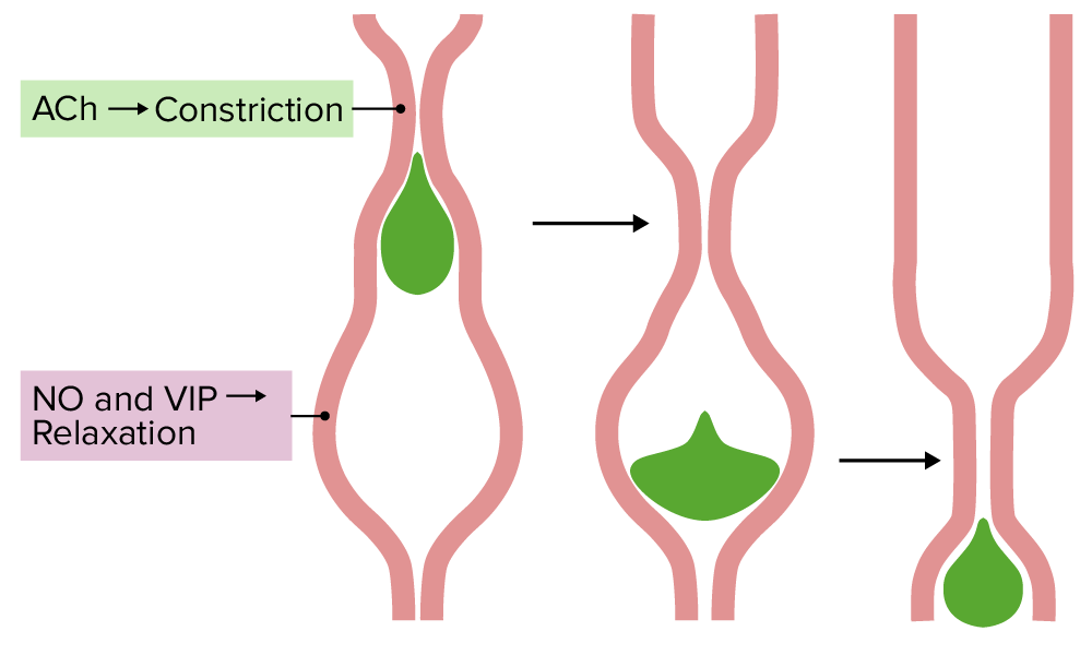
00:02 Hello! Today, we’re going to talk about GI motility. 00:06 We’re going to cover a number of important topics and the first of which is to try to explain how GI motility works. 00:12 And this will include three important aspects including peristalsis, slow waves, and migrating motor complexes. 00:21 Then, what we’re going to do is talk about how swallowing is initiated and controlled. 00:26 And finally, we will explain how gastric emptying works and how this process is regulated and controlled. 00:33 So let’s go back to our GI functions and determine which ones we’re going to cover today. 00:39 And that is excretion, storage, and most importantly, motility. 00:46 To discuss motility, we need to go through a little bit deeper on the various layers of the GI tract. 00:53 These layers go from the mucosal layer, which is the most innermost, to the serosal layer, which is the most outermost. 01:01 So the nerves are going to be controlling the muscular activity, the mucosal cell or layer, then that is the layer of muscle around the mucosal layer. 01:13 Then we have a couple of plexus. 01:15 The Meissner plexus and the submucosal plexus. 01:21 The submucosa, and this is important, this circular layer of muscle. 01:26 And finally, the Auerbach complex, and the longitudinal layer. 01:32 So you notice there were three different layers of muscles in the GI tract. 01:36 And this will be important in being able to squeeze the GI tract together and to be able to push items along this particular tube. 01:47 And of course, the final layer is the serosa. 01:52 Now, how this process works is through an intricate process known as persitalsis. 01:57 Now, peristalsis is a way of coordinating muscle contraction and this is smooth muscle contraction. 02:05 So you’re going to use acetylcholine at the site to cause a contraction. 02:11 We need to relax the area that is in front of that contraction. 02:15 And that is done via nitric oxide or vasoactive intestinal peptide. 02:21 And these are released right where you need to relax that particular portion of the muscle. 02:29 Then, what happens is the food stuff is pushed along because you have a contraction and a relaxation. 02:36 And of course, everything is going to follow to the area of lower pressure. 02:40 This whole process is controlled either by the parasympathetic nervous system or the enteric nervous system depending on if it is centrally driven or if it is a local reflex. 02:51 The central driven is the parasympathetic nervous system and the local reflex is the enteric nervous system. 02:59 Now, besides peristalsis, the other important thing are these sphincters. 03:04 Sphincters will be areas that are constricted during rest and this prevents movement from one area of the GI system to another. 03:13 And what mediates this constriction are enkephalins. 03:17 Often though, vasoactive intestinal peptide will be the relaxation agent for those particular sphincters. 03:25 So when you want to open them, you would give the VIP or nitric oxide some time to be mediated to cause relaxation. 03:35 Now, the other motility issues that need to be discussed besides peristalsis and contraction of sphincters are a lot of the movements are not to just to move things through the GI tract, but rather to mix things up. 03:50 Because we have all these secretions in the GI system. 03:53 What you need to do is make sure they’re well mixed and so the enzymes can work in the appropriate spots.
About the Lecture
The lecture Introduction – Gastrointestinal Motility by Thad Wilson, PhD is from the course Gastrointestinal Physiology.
Included Quiz Questions
Which of the following signaling molecules facilitates the contraction aspect of peristalsis?
- Acetylcholine
- Nitric oxide
- Vasoactive intestinal peptide
- Enkephalin
- Norepinephrine
Which of the following is the inner most layer of the gastrointestinal tract?
- Mucosal layer
- Serosal layer
- Longitudinal muscle layer
- Circular muscle layer
- Nerve plexus layer
Which of the following is an effect of nitric oxide on gastrointestinal motility?
- Sphincter relaxation
- Sphincter constriction
- Gastrin release
- Gastrin inhibition
- Decreased gastric motility
Customer reviews
1,0 of 5 stars
| 5 Stars |
|
0 |
| 4 Stars |
|
0 |
| 3 Stars |
|
0 |
| 2 Stars |
|
0 |
| 1 Star |
|
1 |
In general, I find the lectures provided in Lecture to be of exceptional quality. This one is not only below that standard, but, for me, it fails entirely as an instructive tool especially in regard to the discussion around the first figure. Basic principles to convey information are missing, and when combined together make for a frustrating experience where little to no information is really conveyed. 1. The labels are indicated only as the lecturer speaks. The slide never shows the labels at one time only. (I understand the lecturer wishes to flag the information he is talking about. This could just as easily been done with a figure that shows all of the labels but uses color or boldness to draw our attention to the item under discussion. Better yet, in lieu of highlighting the label, highlighting the actual anatomical item is even more effective). 2. The labels are not adjacent to each other so that the reader looses a visual relation. This is a circular structure with consistent layering, yet the labels jump from side to side for no reason. This is not only distracting but prevents us viewers from easily following the structure along with the discussion. 3. There is no rhyme or reason to the coloring. Connective tissue and muscle are both pink and yellow. This makes it impossible for the viewer to quickly appreciate the structures. Frankly, from this lecture, I have no clue what is where in terms of the layer of the esophagus and the location of each plexus. I ask each of you at Lecturio who are reading this review to see if you are able to grasp this information after reviewing this lecture. In addition to fixing this lecture, it is my hope to hear from Lecturio as to whether the company has in place an editorial team to ensure that basic principals to successfully convey information, as has been done in this lecture, are never missed. After all, we students are paying for a professional service to help educate us, not waste our time. Thank you for your consideration.


