Playlist
Show Playlist
Hide Playlist
Inspection of the Chest – Lung Examination

-
Reference List Physical Examination.pdf
-
Download Lecture Overview
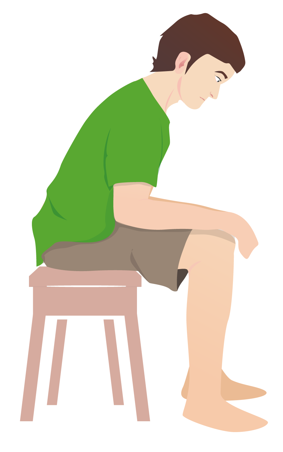
00:01 Next up, we're going to do the respiratory exam and like examination of many systems it starts with inspection, so I don't know about you, but I've taken care of a number of patients who tell me that they're unable to breath and having significant respiratory distress and they can go into great detail about that without even taking a breath. 00:18 In reality, a person who is legitimately having respiratory distress should typically have a problem where they can't say more than a few words at a time. 00:27 So just looking at our patients right now, he looks comfortable and if I ask him to tell me how he was feeling, as long as he can get out a few words, a full sentence, etcetera, then I know that he's not in significant respiratory distress. 00:39 It's worth documenting that in your physical exam, your patient can speak in only one word or three word sentences to really get it that picture very quickly. 00:47 In addition, patients who are in respiratory distress oftentimes use accessory muscles of respiration, so remember that normally, if you're just breathing standard tidal volumes at rest, you'll only need one muscle to do that, and that's your diaphragm, so just looking at our patient relaxing right now, taking normal tidal breaths of inhalation and exhalation, the only muscle that's working is his diaphragm which right now is up, and now it's going down, then it goes up, it's going down to draw air into his chest and then it relaxes. 01:20 And it's actually, for inhalation, that's the only time the diaphragm is doing anything, during exhalation it's simply the elastic recoil of the rib cage that is expelling air from your lungs. 01:32 In contrast, a person who's in respiratory distress is going to use extra muscle groups to help with both inhalation and exhalation, so let's first look at the muscles of inhalation, the accessory muscles of inhalation. 01:46 So what I'm going to have you do, Shaun is I want you to take a full deep breath, as big as you can, go ahead. 01:54 You'll note, you can see right here what happened. 01:55 His external cleidomastoids and his scalene muscles pulled up his clavicles as well as his first rib, and by doing so, he's increasing the amount of space in his chest cavity so he can accommodate more air coming in. 02:09 So that's one of the first sign that you'll see for inhalation in terms of accessory muscle use. 02:14 And now what I'm going to have you do Shaun is completely exhale, completely empty your chest of air. 02:25 Great. So you can see right here he's contracting, you can relax now, he's contracting his abdominal muscles because by contracting his abdominal muscles, he is squeezing his abdominal cavity which pushes the diaphragm upwards and allows extra ability to expel air from the lungs. 02:39 In addition, you may have noticed a subtle change where he leaned forward just a little bit. 02:44 With that was, was his intercostal muscles of his rib cage also contracting which basically just closes the cage to again assist with expelling air from the lungs. 02:55 Other findings that particularly you might see in a person with a COPD exacerbation is pursed lip breathing. 03:04 When patients have emphysema, there's a loss of the architecture that supports the patency of small airways so when they're trying to expel air, the airways are collapsing. 03:15 When you do pursed lip breathing, just basically purse your lips and try and breath out, kind of more like this. With pursed lip breathing, he's creating a back pressure, so called positive end expiratory pressure that basically tents open those smaller airways to allow that air to get out more effectively in the setting of emphysema. 03:42 Okay, you can relax. 03:45 The next thing you might see in a patient with emphysema is the tripoding position. 03:48 So this is known as the tripoding position and the patient is essentially using his arms to prop up his upper thorax which allows him to still keep that rib cage very open but reduces the work of the accessory muscles of respiration in the process. 04:03 So this is another thing that you may find in patients who particularly have emphysema who are in respiratory distress. 04:09 And perhaps the last and perhaps most significant finding that you'll see in a person who is legitimately in respiratory distress is cyanosis and we know this from literature and movies and everything else that the patient gets blue lips, and this is a very real finding with really exceptional likelihood ratios as well. 04:27 So for a person who has an oxygen saturation of less than 80% and you can sort of put this on your finger here, so we'd be looking at his oxygen saturation to tell us what his O2 sat is, but even before we have an O2 sat monitor, I'm just going to basically take a look at his lips. 04:44 The patient with cyanosis they should have a clear bluish discoloration and Shaun can you open your mouth for me? The tongue, stick it out for me, would also be somewhat bluish in color and lifting up your tongue, underneath those mucus membranes beneath the tongue is where you're most likely to see evidence of this bluish discoloration that is evidence of deoxyhemoglobinemia. 05:03 This has a likelihood ratio, a negative likelihood ratio of 0.2 and if you remember from our introductory lecture, a negative likelihood ratio of 0.2 correlates with a change to the pretest probability of 30%.
About the Lecture
The lecture Inspection of the Chest – Lung Examination by Stephen Holt, MD, MS is from the course Examination of Cardiovascular and Respiratory System.
Included Quiz Questions
What sign is seen in a patient with respiratory distress?
- Cannot speak in full sentences without needing to take a breath
- Pacing around the room
- Sitting up straight on the exam table
- Speaking rapidly for several minutes to describe their shortness of breath
- Swallowing saliva more often
What is the "tripod" position seen in a patient with respiratory distress?
- The patient sits or stands leaning forward and supports the upper body with their hands on the knees.
- The patient sits straight up on the exam table with an erect posture.
- The patient needs to lie down on the exam table.
- The patient needs to stand up to breathe comfortably.
- The patient uses neck muscles and abdominal muscles to help with breathing.
Which description indicates a patient with cyanosis?
- A patient with bluish discoloration of the skin or mucous membranes under the tongue
- A patient using neck muscles and abdominal muscles to help with breathing
- A patient sitting, leaning forward to support their upper body with their hands on the knees
- A patient exhaling through tightly pressed lips and inhaling through the nose with the mouth closed
- A patient who cannot speak in full sentences due to respiratory distress
Author of lecture Inspection of the Chest – Lung Examination

Stephen Holt, MD, MS
Customer reviews
5,0 of 5 stars
| 5 Stars |
|
5 |
| 4 Stars |
|
0 |
| 3 Stars |
|
0 |
| 2 Stars |
|
0 |
| 1 Star |
|
0 |
