Playlist
Show Playlist
Hide Playlist
Inguinal Region

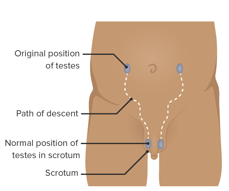
00:00 Inguinal region, the thing, most important bit. 00:06 Do you want to sit down for one minute? Inguinal region, what you need to remember, the inguinal canal, boundaries, contents. 00:16 Now, unfortunately, it is so much of theory. 00:20 I can't explain the inguinal canal. 00:23 So, you have the external oblique coming here, internal oblique that way. 00:27 So, this essentially forms your anterior and superior part. 00:30 Can you get it? It's like a canal, like a tunnel, isn't it? So, your external oblique and the internal oblique essentially forms the superior and the anterior part. 00:41 This part is the inguinal ligament and posterior wall is transversalis fascia. 00:47 This is all you need to know. 00:49 But then when it comes to the medial side, you have the conjoint tendon because that's conjoint tendon between the internal oblique and the external oblique. 00:56 You also have a little bit of the lacunar ligament. 01:00 You need to remember this concept. 01:02 External oblique going this way, internal oblique coming and this is your roof. 01:11 Okay. So, what are the contents of the inguinal canal? So, you have the spermatic cord and in male, ilioinguinal nerve. 01:20 In female, round ligament of the uterus and ilioinguinal nerve. 01:24 So, those are the contents. 01:26 So, coming down here, scrotum, spermatic cord, What are the contents of the spermatic cord? Pampiniform plexus or veins, cremasteric artery, Vas deferens you mean? Vas deferens, yeah. 01:51 Spermatic fascia, internal and external Very good. 01:54 So, the easiest way to remember is rule of threes. 01:57 You have three fasciae, three nerves, three arteries, and three other structures. 02:04 So, the three fasciae are internal spermatic fascia, external spermatic fascia, and cremasteric fascia. 02:12 These three fasciae are essentially the extension of your external oblique, internal oblique, and transverse abdominis. 02:20 These are the extension. 02:22 So, they are called external spermatic fascia, internal spermatic fascia, cremasteric fascia, so three fasciae. 02:28 What are the three arteries? Cremasteric artery. 02:30 Cremasteric artery, testicular artery, and artery to the vas deferens, three arteries. 02:38 Three nerves: genital branch of the genitofemoral nerve. 02:44 Then you have some sympathetic nerves. 02:46 The third one we normally talk about is ilioinguinal nerve, although it’s outside the canal, outside the spermatic cord. 02:55 What are three other structures? Vas deferens, lymphatics, and pampiniform plexus or veins. 03:07 Those are your spermatic cord structures. 03:13 So, that brings us to the femoral triangle, left side, femoral triangle. 03:27 I’ve drawn the femoral artery here. 03:29 What’s here? Femoral vein. 03:34 Here? Nerve. 03:40 Vein, artery, nerve, so, NAV. Nerve, artery, vein... 03:47 Out of this, what is within the femoral sheath and what is outside? No, nerve is outside. Nerve is outside the femoral sheath. 04:01 Artery, vein, and the femoral canal is within the femoral sheath. 04:10 So, artery, vein, femoral canal. 04:16 Significance of femoral canal, femoral hernia strangulates. 04:20 What are the boundaries of the femoral canal? Medially, lacunar ligament. 04:31 Well, here. 04:32 Posteriorly, inguinal ligament, no, no, no, no. 04:42 Hang on a minute. It’s this way. 04:45 It’s coming out this way. Here is inguinal ligament. 04:51 What ligament is this? Pectineal ligament, pectineal ligament. 04:55 What is on the medial side? It's a lacunar ligament or even more part of the conjoint tendon coming off your arch. 05:04 Laterally, you have the femoral vein. 05:06 So, essentially, the point is there is no scope for it to expand. 05:10 It is tight in all the four areas. 05:13 So, the only area it can expand is onto the femoral vein. 05:17 So, that's why it's so dangerous. 05:19 Inguinal ligament, pectineal ligament, or tight ligaments, it doesn’t move at all. 05:26 Then in the middle side is we are so close to the bone, to the pubic symphysis or the tubercle. So, nothing to expand. 05:32 On the lateral side, you have the femoral vein. 05:34 All these are part of the femoral sheath. 05:37 This is outside the femoral sheath. 05:39 What else do you have here? Lymphatics, some lymph nodes, Cloquet's node and the inguinal lymph nodes. 05:47 What muscle is this? Sartorius, adductor? This is adductor magnus. Which part of adductor magnus? Yeah, that’s fine. 06:05 The way the adductor magnus muscle is like this. 06:14 This part is your middle border. This is the lateral border, sartorius. 06:22 These are your contents. 06:25 What is the floor of the femoral triangle? What are the muscles on the floor of the femoral triangle? I'm sure you know this. 06:40 Tell me what happen, how do you do flexion of the hip? It was in the iliacus. Iliacus, psoas, what else? Pectineus, so that’s why you have the pectineal ligament. 06:51 Remember? That's why you have the pectineal ligament at the back because you have the pectineus, iliacus, and psoas, PIP, pectineus, iliacus, and psoas. 07:02 Those are the boundaries, the contents. 07:06 Will you stand up for me, please? Do you mind standing on a chair? Is that too much to ask? No. I'll stand. Sure. 07:15 Thank you. 07:17 I’ll just quickly because otherwise, you won't be able to see from there. 07:22 Basically, we come up to the apex of the triangle here. 07:27 That’s the apex of the triangle. 07:29 This is where the adductor longus, that’s the sartorius and the adductor longus is here. 07:39 This forms the subsartorial canal, also called the Hunter’s canal. 07:43 So, from this point onwards, the femoral artery goes from the anterior compartment to the posterior compartment and begins the popliteal artery. 07:53 Likewise, the femoral vein comes from the posterior compartment to the anterior compartment and forms the femoral vein. 08:01 The femoral nerve comes from here. 08:04 What happens after this femoral nerve? What is the name called? Sensory, yeah. 08:12 What's the name? No. Saphenous nerve. Yeah. 08:18 So, the femoral nerve is a sensory nerve, sorry, is a mixed nerve. 08:23 It's more until there. Beyond this, it becomes sensory. 08:29 It is called the saphenous nerve which accompanies a long saphenous vein. 08:37 So, that's the Hunter's canal. 08:39 What's the nerve supply to the anterior compartment of the leg? Femoral nerve. 08:43 What’s the nerve supply to the middle compartment of the leg? Obturator nerve, okay. 08:47 Do stand down for a minute. I’ll just explain to them. 08:50 You can sit down.
About the Lecture
The lecture Inguinal Region by Stuart Enoch, PhD is from the course Upper Part of the Body Anatomy.
Author of lecture Inguinal Region

Stuart Enoch, PhD
Customer reviews
5,0 of 5 stars
| 5 Stars |
|
5 |
| 4 Stars |
|
0 |
| 3 Stars |
|
0 |
| 2 Stars |
|
0 |
| 1 Star |
|
0 |
