Playlist
Show Playlist
Hide Playlist
Hydrocephalus and Masses

-
Slides Hydrocephalus and Masses.pdf
-
Download Lecture Overview
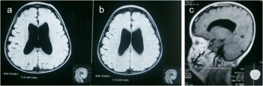
00:01 So let's talk a little bit about intracranial abnormalities. 00:04 First we'll discuss hydrocephalus. 00:09 Hydrocephalus is the increase in volume of CSF that results in expansion of the ventricular system. 00:15 This could be caused by several different reasons. 00:18 You can have overproduction of the CSF. 00:20 You can have decreased absorption of the CSF or you can have obstruction of CSF flow and this results in disproportionate dilatation of the ventricles in comparison to the sulci when you take a look at a scan of the brain. 00:33 There two major types of hydrocephalus, there's communicating and there's non-communicating. 00:39 Communicating hydrocephalus is caused by a mismatch of CSF production and absorption. Usually this is caused by something that inhibits absorption at the level of the arachnoid villi, such as metastatic deposits. 00:51 Non-communicating is usually caused by physically obstructing lesions that block the outflow of CSF. This can be treated surgically by removing the obstructing lesion. 01:02 So let's take a look at this case. This is an axial CT image through the brain and you can see if this patient has normal appearing sulci. 01:10 They don't look like they're dilated. 01:12 However, the ventricles do look like they're significantly dilated and so these are dilated out of proportion with respect to the sulci. 01:20 This is an example of a communicating hydrocephalus with disproportionate enlargement of the ventricles with respect to the sulci. 01:28 So when we discuss masses within the brain, we wanna be able to determine whether they are intra-axial or extra axial. 01:35 Intra-axial or intracranial mean that they're within the substance of the brain. 01:40 Extra-axial means that they're outside of the brain but within the skull. 01:44 So Gliomas are the most common primary intracranial malignancy. 01:50 Intracranial metastasis are equally common. 01:53 Some of the CT characteristics of intracranial masses include a hypodense or isodense mass that seen on CT. 02:01 It can cause surrounding edema and it enhances with contrast administration. 02:07 So there are two major types of cerebral edema. 02:10 There's vasogenic edema, which is extracellular fluid accumulation that's caused primarily by a mass or an infection. 02:16 And this is localized to the white matter of the brain. 02:19 There's also cytotoxic edema which is cellular edema and that's associated with cell death and that's usually caused by ischemia or infarction, that affects both the gray and the white matter and it results in loss of gray/white differentiation. 02:33 So here's an example, we have two different axial, non-contrast CT images of the brain. 02:39 So which of these represent vasogenic edema and which one represents cytotoxic edema? So if you think back to the description, the left image does demonstrates vasogenic edema in a patient that has a glioma. 02:59 The right image demonstrates cytotoxic edema in a patient that has a large right infarction. 03:05 Both of them have subfalcine herniation. 03:09 So let's take a better look. This image right here is vasogenic edema because it involves primarily the white matter. 03:16 It has sparing of the gray matter which we see here. 03:19 On the right, we have involvement of both the gray and the white matter indicating that this one is cytotoxic edema and if you take a look at the midline, the midline is moved over to the left meaning that this is subfalcine herniation. 03:36 This happens on this image as well where the midline is moved over to the left. 03:42 So meningioma is the most common extra-axial mass lesion. 03:46 Usually these are very slow growing and benign and they often don't need to be surgically removed. 03:51 This is an axial CT image of the brain which demonstrates a very high density mass which represents a meningioma. 04:00 So meningioma is typically are hyper-dense on CT. 04:04 They often contain calcifications which leads to their increase in density and they have significant enhancement on post-contrast images. 04:12 They have what's called a dural tail which is a small piece of the mass extending along the dura and they can cause adjacent vasogenic edema because of there mass effect even though they are extra-axial in location. 04:24 So the dural tail is a portion of the mass that extends along the dura and this helps us identify its extra-axial location. 04:31 Remember, one of the key things that you have to do when you see a mass within the head is determined whether it's inter-axial or extra-axial. 04:38 So something like a dural tail will help you with that.
About the Lecture
The lecture Hydrocephalus and Masses by Hetal Verma, MD is from the course Neuroradiology.
Included Quiz Questions
What is TRUE regarding cytotoxic edema?
- It is seen with acute infarction.
- It is extracellular fluid accumulation.
- It is associated with masses.
- It is localized to the white matter.
- It preserves gray/white differentiation.
What is TRUE regarding hydrocephalus?
- It results in an expansion of the ventricular system.
- It is due to decreased production of the CSF.
- It is due to increasing the cortex mass.
- There are 3 major types; communicating, non-communicating, and mixed.
- It is due to increased absorption of CSF.
Which of the following is a slow-growing benign brain tumor?
- Meningioma
- Glioblastoma multiforme
- Astrocytoma
- Oligodendroglioma
- Ependymoma
Author of lecture Hydrocephalus and Masses

Hetal Verma, MD
Customer reviews
5,0 of 5 stars
| 5 Stars |
|
5 |
| 4 Stars |
|
0 |
| 3 Stars |
|
0 |
| 2 Stars |
|
0 |
| 1 Star |
|
0 |
