Playlist
Show Playlist
Hide Playlist
Hydatidiform Mole

-
Slides PregnancyComplications Female ReproductivePathology.pdf
-
Reference List Pathology.pdf
-
Download Lecture Overview
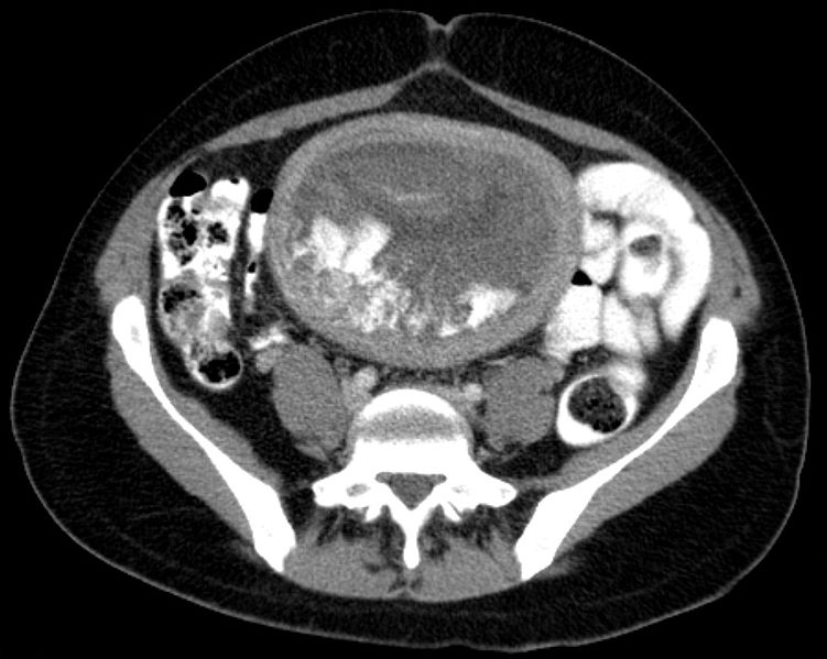
00:01 Our next topic is hydatidiform mole. 00:05 What exactly is the mole? And what are we referring to? Or big picture? Our section here is placental pathologies. 00:14 That’s exactly what’s going on. 00:15 It’s the placenta that is going to then – Well, let me show you different characteristics. 00:21 We’ll take a look at partial and complete mole. 00:24 The mole refers to the fact of how much of the placenta is being affected and whether or not you’ll actually find fetal remnants within the placenta. 00:36 Presents in fourth to fifth month of pregnancy, so relatively early in pregnancy. 00:43 There’d be vaginal bleeding in both partial and complete as we shall see. 00:48 Uterus that is larger than expected for duration of pregnancy. 00:51 The gestational age in which you would expect full uterine size size is much, much greater in hydatidiform mole. 00:58 This particular picture that you’re seeing here would be that in which all of the chorionic villi have now become inflamed or dilated. 01:06 All, every single finger-like projection of the chorionic villi, which is part of the placenta, has now become dilated. 01:15 If every single chorionic villi has then become dilated, then you have to call this a complete hydatidiform mole. 01:23 What is the name of the cancer that may then take place of the placenta? That is called choriocarcinoma. 01:31 So there are two types that we’ll take a look at with hydatidiform mole, complete and partial. 01:35 The “C” in complete has a greater risk of going onto “C”, choriocarcinoma. 01:41 Every single chorionic villi has now become dilated. 01:46 What else do you want to keep in mind? The chromosomes. 01:50 So what’s a normal chromosome? Obviously, either 46 XX or 46 XY. 01:57 If it’s a complete mole, you’ll find that there is actually normal chromosomes, 46 XX or 46 XY. 02:04 How is that possible? Hold on for a second. 02:07 Here, because we have such abnormalities of the placenta, you’re going to find extreme, extreme high levels of beta hCG. 02:17 If you were to then take a look at this uterus and placenta, with every single chorionic villi that’s become inflamed -- Well, you might have heard of the term snowstorm appearance. 02:28 That’s exactly what you’d find on your ultrasound, that the dilated chorionic villi looks like a snowstorm upon your ultrasound image. 02:38 Hydatidiform mole, if you take a look at the ultrasound, it looks like a snowstorm. 02:42 I showed you a gross picture and ultrasound. 02:45 Histology might then show you cystic swelling, swelling, swelling of the chorionic villi, especially the complete hydatidiform mole. 02:54 And the complete is the one that has a greater risk of going onto choriocarcinoma. 02:58 In any case, you would find extremely high levels of beta hCG. 03:02 The two different types of moles that we have to know in greater detail. 03:06 Let’s begin. 03:08 Complete mole: Can you tell me a few things about this before we begin? So that you can predict and you get a better idea, reinforcement. 03:14 C – Complete. 03:15 Greater risk of going onto choriocarcinoma. 03:18 C – Complete. What does that mean to you? Every single chorionic villi has now become dilated. 03:22 I’ve now emphasized that over and over again. Why? Why am I making that a big deal? Well, I’ll tell you now. 03:30 Choriocarcinoma, those chorionic villi, they’re absent. 03:34 So you absolutely want to pay attention to description of the chorionic villi. 03:39 If they’re telling you in a stem of a question that every single chorionic villi is inflamed or dilated, that’s a complete mole and take a look at the chromosome, 46 XX. 03:50 “Dr. Raj, that looks perfectly normal.” Yes. 03:53 On appearance, it seems perfectly normal, but that is deceiving, isn’t it? Because you take a look inside the placenta, it’s empty. 04:03 There’s no fetus. 04:04 “So Dr. Raj, how can you have 46 XX and have no fetus?” Because it’s always a man’s fault. 04:09 Always. Know that. 04:11 What do you mean? Well the sperm which is 23 haploid, 23 X or 23 Y. 04:18 It may then double. 04:19 If all that you have is participation of the sperm with no female participation, how in the world are you supposed to have a fetus? You don’t. 04:27 Empty. 04:28 So you have a sperm that will duplicate or dispermy, you’ve heard of. 04:33 That means that two sperms will literally come on and try to fertilize, but that makes no sense either if you don’t have participation of the fetus – excuse me, the female. 04:43 So therefore, you will have 46 XX commonly or maybe perhaps 46 XY. 04:49 However, there are no fetal parts inside the placenta. 04:54 Complete. Take the O in complete. 04:57 Use it to your advantage, for no fetal parts. 05:01 Take the “C” in complete. 05:03 Use that to your advantage, because this will give rise to choriocarcinoma. 05:07 On the other side, we have partial mole. 05:10 You take the “part” in partial and you’ll find parts of the fetus in the placenta. 05:17 So you will actually find parts. 05:19 Now, the description, chromosome Y, this is called triploid. 05:24 What does that mean? Well, instead of 46 XX, it could be something where you have your 69 XXX, 69 XXY. 05:35 Triploid, triploid, triploid. 05:36 Instead of having diploid XX, you have XXY or XXX. You get the point. 05:41 69, 23, 23, 23. 05:44 How is this possible? Well, not only is it the mans fault, but now there’s going to female participation. 05:52 So you’ll have an egg and it will contribute and there will two sperms that will contribute. 05:59 What do you get total? A triploid. 06:02 However, with this type of participation, you’re not going to find a normal fetal development in the placenta. 06:08 So you’ll find fetal parts, fetal parts, that’s huge. 06:12 Once again here, partial mole, you don’t have dilation of every single chorionic villi. 06:17 And the chance of going onto choriocarcinoma, much less than what it is for complete. 06:23 Now that you have a full picture, let’s go onto description. 06:26 Complete mole, can you see as to how quickly now it makes sense? All villi in the complete mole completely are edematous and dilated. 06:36 There’s going to be complete trophoblastic proliferation. 06:39 That’s for complete. 06:41 What about the partial? Some villi edematous and partially surrounded by trophoblastic proliferation. 06:48 Tell me about the fetal content in your placenta. 06:51 Complete - Zip. O. No. 06:53 Partial - Part. 06:56 Complete, no. 06:57 Partial, fetal parts. 06:59 Complete mole, higher risk of choriocarcinoma. 07:02 Partial mole, I’m not saying there isn’t a risk, but there’s a lower risk of choriocarcinoma.
About the Lecture
The lecture Hydatidiform Mole by Carlo Raj, MD is from the course Pregnancy Complications.
Included Quiz Questions
Which of the following is LEAST likely in a patient presenting with a hydatidiform mole?
- Presentation in the 2nd to 3rd month of pregnancy
- Vaginal bleeding
- Extremely high beta-hCG level
- Passage of grape-like tissue from the vagina
- Uterus larger than expected for gestational age
What pattern is seen on an ultrasound scan of a patient with a hydatidiform mole?
- Snowstorm pattern
- Double decidual sac
- Heterogenous uterus
- Non-homogenous echoic pattern
- Pseudosac
Which of the following is associated with a complete hydatidiform mole?
- High risk for choriocarcinoma
- Triploid chromosome
- Some villi that are edematous
- Fetal parts
- 2 sperm and 1 egg
Which of the following is associated with a partial hydatidiform mole?
- Triploid chromosome
- High risk for choriocarcinoma
- All chorionic villi are edematous
- No fetal parts
- 2 sperm with no egg
Author of lecture Hydatidiform Mole

Carlo Raj, MD
Customer reviews
5,0 of 5 stars
| 5 Stars |
|
2 |
| 4 Stars |
|
0 |
| 3 Stars |
|
0 |
| 2 Stars |
|
0 |
| 1 Star |
|
0 |
cool lecturer great and simple explanation. Helps ground you on the basics real fast.
it was well presented, very easy to understand. Godd job Dr Raj
