Playlist
Show Playlist
Hide Playlist
Histology of the Three Muscle Types, and Their Responses to Stress and Injury

-
Slides 06 Types of Tissues Meyer.pdf
-
Reference List Histology.pdf
-
Download Lecture Overview
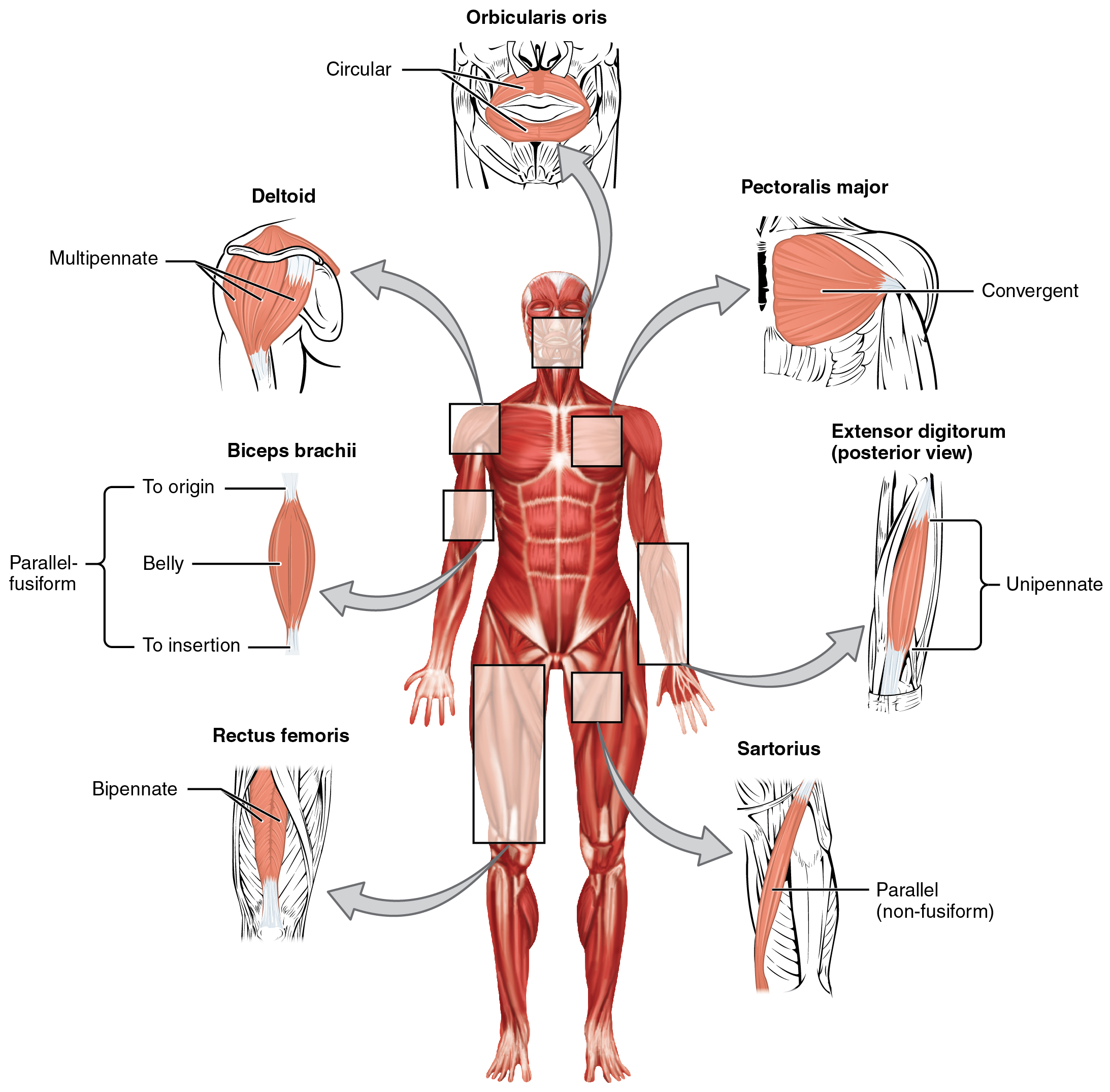
00:00 Well let us look and see how muscle can repair itself after injury. Remember this slide, we explained the components of muscle earlier on the lecture. Focus on the nucleus or said earlier that muscle fibres are multinucleated. 00:18 Well, some of these nuclei are in fact stem cells. They are called satellite cells. 00:26 You cannot tell the difference between the satellite cells and the true skeletal muscle nucleus, but these satellite cells sit on the periphery of the skeletal muscle fibre and they have a nucleus also. As I've said you cannot tell the difference between the nucleus of the satellite cells and the true nucleus of the skeletal muscle fibre. But if skeletal muscle is damaged, these satellite cells can lay down more contractile proteins. The muscle cell cannot divide, it is fully differentiated. It can only rely on the satellite cells to repair the damaged fibre to lay down more contractile proteins that will allow the fibre to get thicker and thicker. If you do weightlifting exercises and you increase muscle mass, you do not get more muscle cells because muscle cells cannot divide, at least the skeletal muscles cannot divide. The muscle fibre gets bigger, thicker, hypertrophies because of these satellite cells laying down more contractile proteins. I want to briefly just explain to you the difference between striated skeletal muscle and striated cardiac muscle. I am going to deal with cardiac muscle in more detail when we look at the cardiovascular system. 01:56 But cardiac muscle fibres are smaller and they are joined end to end at special locations called intercalated discs. These consists of very tight adherent junctions and also gap junctions. And that allows you wave of contraction or depolarization to pass along the length of the cells along, the length of the cardiac muscle components, and therefore bring about a sequential contraction of the cardiac muscle that is important in pumping blood out of the heart. Cardiac muscle is striated, but it has a central nucleus and only one nucleus. So the fact that it has a central nucleus and only one nucleus and it has these intercalated discs is a sort of specialized structural details that separate it from a normal striated skeletal muscle fibre. Let us quickly look at the smooth muscle fibre. They are not striated. They're joined together by gap junctions, and those gap junctions allow again the wave of contraction or the wave of depolarization to move along the length of the massive smooth muscle and bring about again a sequential contraction. 03:23 They have one central nucleus like cardiac muscle, but they do not have striations. And that again is enough criteria for you to now to be able to distinguish between skeletal muscle fibres, cardiac muscle fibres and smooth muscle fibres. Smooth muscle can respond to injury. 03:43 In the case of the artery wall, smooth muscle cells can divide and the wall can get thicker and thicker if need be. In the endometrium of the uterus, the muscle cells can divide and also hypertrophy. We see that during pregnancy. And the muscular external layers of smooth muscle in the gut tube can also divide and hypertrophy. So smooth muscle has the ability to grow and repair because they can divide. They can undergo regular mitotic activity. 04:16 While in summary then, make sure you can distinguish the three muscle types, skeletal, cardiac and smooth muscle and those that are striated and those that are not. Make sure you understand particularly with skeletal muscle about innervation of these muscle fibres. Understand the wrappings around the skeletal muscle and how those wrappings come together to form a myotendinous junction. Make sure you are aware of the different muscle fibre types, the red, the intermediate and the light whiter pink fibres. And also understand the structure of the sarcomere and be able to identify the sarcomere in the skeletal muscle fibre. And finally be aware of the function of both the neuromuscular spindle and the Golgi tendon organ. They allow us to perceive our position in space, the position of our limbs because these receptor organs tell the central nervous system about the stretch and length change in muscle and also about muscle tension. So thank you for listening to this lecture. 05:34 I hope you have enjoyed learning a little bit about skeletal muscle in particular and I hope you look forward to learning more about smooth muscle and cardiac muscle when I deal with those very special muscles when we look at the cardiovascular system and also when we look at other parts of the organ systems and look at the role of smooth muscle in those systems. So once again thank you very much for listening to this lecture.
About the Lecture
The lecture Histology of the Three Muscle Types, and Their Responses to Stress and Injury by Geoffrey Meyer, PhD is from the course Muscle Tissue.
Included Quiz Questions
Multinucleated muscle fibers are predominant in which of the following muscles?
- Sartorius muscle
- Muscles in the wall of the jejunum
- Atrial myocytes
- Muscles in the wall of the stomach
- Muscles of the trachea
Which of the following is NOT a feature of smooth muscle fibers?
- Striated myofibers
- Uninucleated myofibers
- They are under autonomic control.
- They are involuntary muscles.
- The myofibers are spindle-shaped.
Which of the following statements regarding a fully differentiated skeletal muscle cell is correct?
- It is incapable of cell division.
- It may continue to differentiate into a satellite cell.
- It usually undergoes apoptosis after completing differentiation.
- It remains in the cell cycle.
- More actin than myosin is produced after differentiation.
Author of lecture Histology of the Three Muscle Types, and Their Responses to Stress and Injury

Geoffrey Meyer, PhD
Customer reviews
5,0 of 5 stars
| 5 Stars |
|
3 |
| 4 Stars |
|
0 |
| 3 Stars |
|
0 |
| 2 Stars |
|
0 |
| 1 Star |
|
0 |
He is great, I have leant a lot from his easy and smooth teaching method Thanks
i enjoyed the explanations , especially those of the z line , m line, light & dark bands
very good lecturer also listen?ng at 1.25x speed works well. I recommend it.
