Playlist
Show Playlist
Hide Playlist
How Are Exocrine Glands Classified and Named?

-
Slides 02 Types of Tissues Meyer.pdf
-
Reference List Histology.pdf
-
Download Lecture Overview
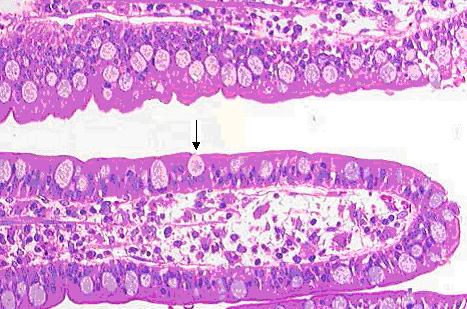
00:01 Now there are different ways in which exocrine glands are named or classified. First of all, if you look at the epithelial surface shown on the left-hand slide, the image of an epithelium in the gut, you can see single glands or single secretory cells called goblet cells that look like a wine goblet. They are shaped like a wine goblet and these are called goblet cells. 00:33 They secrete mucous. There are examples of a unicellular gland, a single cell sitting in an epithelial surface or on its own. Or there are clusters of these cells all the way along the epithelial surface. If you look at the image on the right-hand side, this is an example of a multicellular gland. Here is a sheet of epithelial cells that are secretory, forming a number of layers. And if you look very carefully at this stomach mucosa, these layers of epithelial secretory cells, you can see that they stain differently. 01:16 The surface ones are very clear staining. In the middle, you can see some cells that have a pink, reddish stain and down the base of these epithelial layers of cells, you can see bluish stained cells. And that indicates that in this sheet of secretory epithelial cells, there are three different sources of secretory products. So that is an example of a multicellular gland. 01:50 Now I want you to remember the word mucosa, that I have shown you on the slides, stomach mucosa. The word mucosa is a word or term I am going to use in a number of lectures, particularly when we look at the organ system. So please remember that word because later on towards the end of the lecture, I am going to explain what a mucosa is. I won't do it now because it is more important and more appropriate towards the end, but just remember that word mucosa. Okay, these multicellular glands can get far more complex than just a sheet of cells you saw in that stomach mucosa. They can acquire a duct rather than secreting onto the surface like a goblet cell or like these cells in the stomach mucosa during development. 02:43 When the epithelium invaginates into the underlying lamina propria, the portion that connects the invaginated epithelial cells to the surface is retained, and that forms a conduit or a duct. The deeper epithelial cells that have invaginated into the lamina propria become the secretory cells, the busy cells that make the secretion products. And so we can further classify multicellular glands that're having ducts. And if the ducts don't branch, we call it a simple gland. But if the duct branches like you see in the three diagrams done in the bottom of this slide, we call it a compound gland. Now the classification can go further if you look at the nature or the shape or the structure of the secretory product and I am not going to go through the names listed here and I certainly do not want to emphasize these names or I certainly would not expect you to be able to recall these names, because in reality when you look at sections through multicellular glands, it is very difficult to appreciate the three-dimensional structure and therefore we have to name the glands like you see listed here. It is very different though to needing to remember the why and which epithelia are named and classified.
About the Lecture
The lecture How Are Exocrine Glands Classified and Named? by Geoffrey Meyer, PhD is from the course Epithelial Tissue.
Included Quiz Questions
Which of the following is a unicellular gland?
- Goblet cell
- Mammary gland
- Sebaceous gland
- Sweat gland
- Compound gland
What do goblet cells mainly produce?
- Mucus
- Serous
- Sweat
- Hormones
- Sebum
Which of the following statements about exocrine glands is INCORRECT?
- They secrete their products directly into the bloodstream.
- They may be unicellular or multicellular.
- An example of a unicellular gland is the goblet cell.
- Multicellular exocrine glands may have complex arrangements.
- Multicellular exocrine glands may have more than one secretory cell type.
Author of lecture How Are Exocrine Glands Classified and Named?

Geoffrey Meyer, PhD
Customer reviews
5,0 of 5 stars
| 5 Stars |
|
5 |
| 4 Stars |
|
0 |
| 3 Stars |
|
0 |
| 2 Stars |
|
0 |
| 1 Star |
|
0 |
