Playlist
Show Playlist
Hide Playlist
Hereditary Spherocytosis (HS): Pathogenesis

-
Slides Hereditary Spherocytosis.pdf
-
Reference List Pathology.pdf
-
Download Lecture Overview
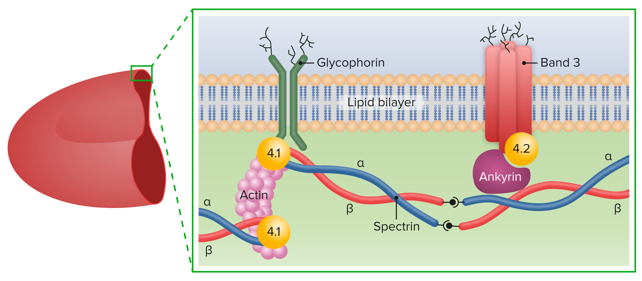
00:01 So now, there are membrane fragments that are being lost. 00:04 You are then going to lose that biconcave shape and you become a sphere. 00:08 Now, that you have a misshapen RBC, do you remember that picture in our discussion where we’re talking about a spherocyte? And inside the spleen, you had these splenic macrophages and remember your guests were saying, “Oh, you really shouldn’t have brought us a spherocyte because we just love it." And so you have a splenic macrophage that is just going to go crazy. 00:31 And eat these spherocytes. 00:32 So therefore, what kind of hemolysis is this? Splenic, extravascular hemolysis. 00:39 So therefore, you’re patient is going to have which one of these? Significant hemoglobinuria or significant jaundice? Significant jaundice. 00:50 Now, let’s take a look at what we have here. 00:54 Mutations in ankyrin or spectrin, I do want you to know two membrane components. 01:00 The first one that I showed you especially was spectrin, but think of this as being an anchor. 01:04 There’s a protein component called ankyrin. 01:08 And if this also becomes lost, then you have as you seen in the picture here, a perfectly, perfectly formed spherocyte in which the central pallor has been lost. 01:18 Now, granted, there are some RBCs in this picture in which there are central pallor, okay, so you will have a mixed picture. 01:26 You can't say that “Oh well, the entire picture is going to be all spherocyte." That’s not practical. 01:32 But you have enough RBCs here that you’re seeing in which you should be able to come up with a diagnosis of at least two actually. 01:41 Are you ready? So two differentials. 01:43 “So Dr. Raj, you’re telling me that there are two differentials or anemias that then appear where the RBC appears as being a spherocyte?” Correct. 01:50 The two major ones are going to be either HS, hereditary spherocytosis, the other one we’ll take a look at and that you must be very familiar with is the warm type of autoimmune hemolytic anemia. 02:02 I will mention that once again and again and again until we get these differentials down. 02:09 So how can you tell the difference between the two? Is this an autoimmune disease, hereditary spherocytosis? No. 02:16 So therefore anything that you would do in terms of your testing for immunity or autoimmunity is something called a Coombs test. 02:24 And that Coombs test is going to be negative for hereditary spherocytosis. 02:31 Where the antihuman globulin test or the Coombs test will be positive for warm type of autoimmune hemolytic anemia. 02:40 Keep that in mind as we move forward. 02:40 I cannot just give you something and not have you realize that students can get this confused with other conditions. 02:48 Now, let’s now take a look at the involvement of the spleen, but then where am I? This is the gallbladder. 02:57 So you connect the splenic destruction of your bilirubin or your hemoglobin to what we’re seeing here in the gallbladder. 03:05 This is cholelithiasis, the type of stones that you’re seeing here, don’t they appear rather black and pigmented? Yes. 03:13 So it cannot be cholesterol, obviously. 03:16 So don’t ever choose that. 03:16 If it’s extravascular hemolysis, please, this is pigment stones made up of bilirubin so they’re black and pigmented. 03:24 This is a sign for extravascular hemolysis. 03:26 Now, in addition, what else may happen? Well, you also have splenomegaly, so you want to keep that in mid. 03:33 And an important test is this. 03:37 I asked you earlier about that osmotic pressure within that concentrated RBC that I showed you a picture of earlier. 03:43 So if you have increased concentration of RBC and you put this into a saline solution, in other words, you’re putting this into a more hypotonic solution, then what can you imagine and what can you think, what’s the behavior of this water going to do? You can predict that the water is going to go into the concentrated spherocyte. 04:06 What do you call this? The osmotic fragility test. 04:09 So what is going to happen to size of the cell? It is then going to increase in size. 04:16 Now, that’s called the osmotic fragility test and with the spleen being enlarged, you are thinking about doing what? Maybe perhaps a splenectomy. 04:25 And whenever you are compromising the function of the spleen, what kind of organisms are you worried about? You’re worried about this encapsulated organisms especially Streptococcus pneumoniae. 04:35 You also Haemophilus influenzae, Klebsiella pneumoniae. 04:37 So you have a bunch there. 04:38 And then what we see here is calcium bilirubinate and this is increased conjugated bilirubin in the bile. 04:45 Conjugated bilirubin.
About the Lecture
The lecture Hereditary Spherocytosis (HS): Pathogenesis by Carlo Raj, MD is from the course Hemolytic Anemia – Red Blood Cell Pathology (RBC).
Included Quiz Questions
Which of the following types of conditions is most likely associated with spherocytes on the peripheral blood film?
- Autoimmune hemolytic anemia
- Disseminated intravascular coagulation
- Sickle cell anemia
- Beta thalassemia
- Iron deficiency anemia
Which of the following test results is a differentiating factor betweeen autoimmune hemolytic anemia and hereditary spherocytosis?
- Negative Coombs test in hereditary spherocytosis
- Negative Coombs test in autoimmune hemolytic anemia
- Negative osmotic fragility test in hereditary spherocytosis
- Positive osmotic fragility test in autoimmune hemolytic anemia
- Positive Coombs test in hereditary spherocytosis
Which of the following tests is most likely to confirm a diagnosis of hereditary spherocytosis?
- Osmotic fragility test
- Coombs test
- Metabisulfate screen
- Ham test
- Sucrose test
Which of the following is the best interpretation of "increased osmotic fragility of red blood cells in hypotonic saline"?
- Decreased capacity of spherocytes to expand
- Increased capacity of spherocytes to expand
- Decreased capacity of normal erythrocytes to expand
- Increased capacity of normal erythrocytes to expand
Author of lecture Hereditary Spherocytosis (HS): Pathogenesis

Carlo Raj, MD
Customer reviews
5,0 of 5 stars
| 5 Stars |
|
5 |
| 4 Stars |
|
0 |
| 3 Stars |
|
0 |
| 2 Stars |
|
0 |
| 1 Star |
|
0 |
