Playlist
Show Playlist
Hide Playlist
Hereditary Spherocytosis (HS): Etiology

-
Slides Hereditary Spherocytosis.pdf
-
Reference List Pathology.pdf
-
Download Lecture Overview
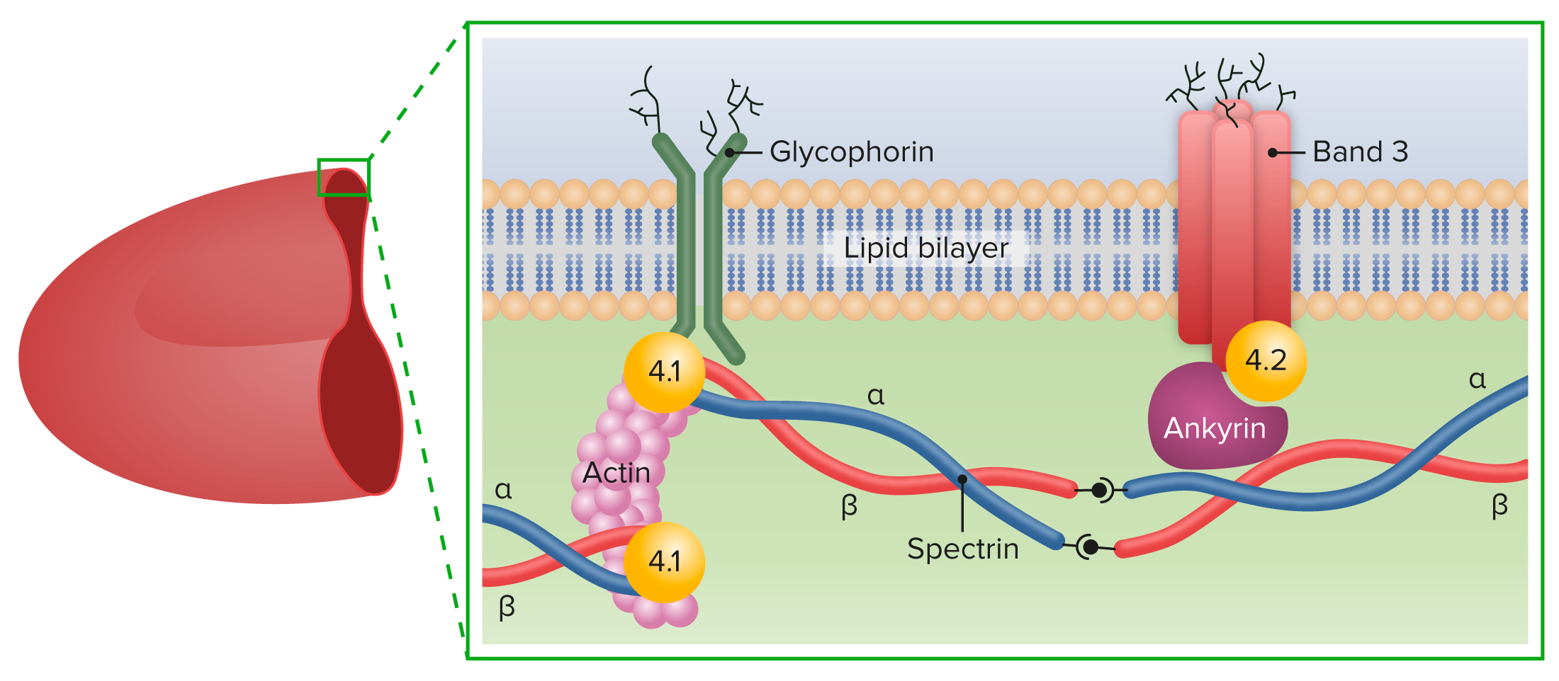
00:01 Let’s take a look at hereditary spherocytosis. 00:05 HS is what I’m going to refer to this as. 00:08 Hereditary spherocytosis is a membrane issue and the name pretty much tells you everything that you need to know. 00:15 Where am I in terms of your category? You are normocytic anemia, you’re looking at MCV between 80 to 100. 00:24 So what then happens when there’s enough membrane loss? Now, in your mind, conceptually work with me here. 00:31 And a normal RBC is biconcave, whatever that means. 00:36 And point being is it has central pallor, right? And so therefore, if you start losing parts of the membrane, what then happens? The membrane is then going to become more and more of a spherocyte. 00:49 It loses its "biconcave shape." What then happens to the central pallor? It’s no longer present. 00:57 So it’s highly concentrated. 01:01 Now you give me another test in which it then tells you that you have a highly concentrated RBC, where the RBCs homogenously reddened or are hyperchromic. 01:14 MCHC. 01:17 Clear? So we have two major RBC index that we’ll take a look at. 01:22 Number one, it’s a fact that, well, your normocytic is where we are, 80 to 100. 01:26 Number two, in terms of reticulocytosis, you expect your reticulocyte production index to be increased or decreased here, please? Increased. 01:36 And we find MCHC because of increased hemoglobin concentration to be high as well. 01:42 So what is this important membrane component that’s missing in HS? Spectrin, spectrin, spectrin. 01:49 You also have ankyrin as well, but spectrin is a major, major membrane component. 01:54 Now, those of you that are interested in going into hematology, you’ll be dealing with and spending a lot of time with that RBC membrane for various reasons. 02:04 There are all of these antigens. 02:06 You all know about D antigen, right? But you have all of these antigens and membrane components that you’ll have to know about. 02:13 At this point, I would know that spectrin is becoming lost, resulting in an RBC that looks like a sphere, spherocyte. 02:23 Next, may I ask you a question? If more that you have concentration increasing within the RBC, then tell me about its osmotic pressure inside the RBC, increased or decreased? Keep that in mind. 02:38 Let’s move on.
About the Lecture
The lecture Hereditary Spherocytosis (HS): Etiology by Carlo Raj, MD is from the course Hemolytic Anemia – Red Blood Cell Pathology (RBC).
Included Quiz Questions
Hereditary spherocytosis is caused by mutations in the gene that codes for which erythrocyte protein?
- Spectrin
- Globin
- Hepcidin
- Haptoglobin
- Transferrin
Which morphological characteristic on the peripheral blood smear distinguishes spherocytes from normal erythrocytes?
- Loss of central pallor
- Spherocytes are indistinguishable from normal erythrocytes on the peripheral blood smear.
- Increased surface area
- Convex shape
- Hypochromia
Author of lecture Hereditary Spherocytosis (HS): Etiology

Carlo Raj, MD
Customer reviews
5,0 of 5 stars
| 5 Stars |
|
5 |
| 4 Stars |
|
0 |
| 3 Stars |
|
0 |
| 2 Stars |
|
0 |
| 1 Star |
|
0 |
