Playlist
Show Playlist
Hide Playlist
Hepatic Cirrhosis

-
Slides Hepatic Cirrhosis.pdf
-
Download Lecture Overview
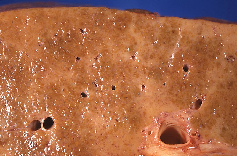
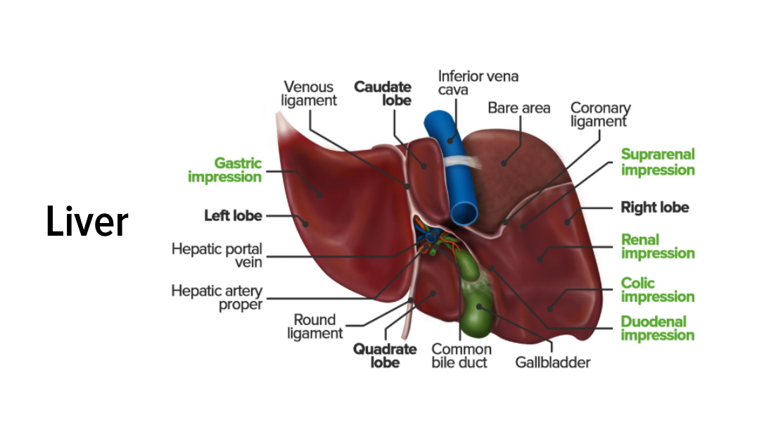
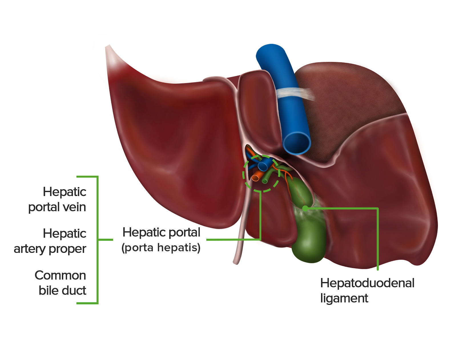
00:01 So in this lecture we'll be talking about hepatic cirrhosis and the imaging findings associated with it. 00:06 What is hepatic cirrhosis? It is chronic liver disease that's characterized by diffuse fibrosis. 00:13 You can see a pathologic picture of hepatic fibrosis and you can see that the entire liver looks slightly abnormal, you have little nodules and the entire liver looks just a little bit shrunken and nodular. 00:25 Some of the common causes of hepatic cirrhosis include chronic hepatitis C which is one of the most common causes worldwide, alcoholic liver disease which is also one of the most common causes of hepatic cirrhosis, hepatic steatosis also called non-alcoholic liver disease, chronic hepatitis B, and occasionally, autoimmune or metabolic conditions which are a lot more rare. 00:51 Some of the complications associated with hepatic cirrhosis include portal hypertension, ascites or free fluid within the abdomen, pleural effusion or fluid within the lung, hepatic failure, renal failure, hepatocellular carcinoma, and possibly, death. 01:11 So, what exactly is portal hypertension? It's elevated pressure within the portal venous system. 01:18 So this diagram here shows you what the portal venous system looks like. 01:22 Here's the portal vein right here and here is the superior mesenteric vein coming down. 01:26 So this entire venous system becomes engorged in portal hypertension. 01:30 Some of the imaging features of portal hypertension include esophageal or splenic varices, reversal of flow or thrombosis of the portal vein, you can have a patent paraumbilical vein, you can have an enlarged spleen or splenomegaly, gallbladder wall thickening, and you can also have stomach wall and small bowel or colon thickening. 01:51 So let's look at some of the contour changes associated with hepatic cirrhosis. 01:57 As we saw on the pathologic picture, even on the radiological picture, you can see a lot of nodularity to the contour of the liver, and this is really one of the earliest signs of hepatic cirrhosis. 02:09 The liver also looks somewhat shrunken; regenerative nodules or nodular proliferation of the liver parenchyma can also be seen but it's hard to appreciate on a CT. 02:18 This is usually better appreciated on an MRI. 02:21 Ascites is also a very common finding and you can see here surrounding the liver a low density fluid collection representing free fluid in the abdomen or ascites. 02:30 So let's discuss other findings of hepatic cirrhosis. 02:38 What do you see on this image? Do you see the abnormality pointed out by the arrows? What do you think that represents? So this is actually an example of a small pleural effusion which is the low density that's back here and adjacent to compressive atelectasis or atelectasis of the lung caused by compression by the pleural effusion, and this is a very common finding of hepatic cirrhosis. 03:07 What kind of ultrasound of the liver is this? This right here is all liver parenchyma, and then what do these colors represent? So this is actually a Doppler ultrasound. 03:27 It's a color Doppler ultrasound of the portal vein and Doppler demonstrates flow within the vessels. 03:34 So this actually shows that there is flow within all of these vessels but there is no flow within the main portal vein right here. 03:41 This indicates portal vein thrombosis which is also a complication of hepatic cirrhosis. 03:46 This is an example of splenomegaly. 03:52 The spleen should be less than 12 cm in longitudinal dimension. 03:55 This is usually best measured on ultrasound but it can also be extrapolated on a CT. 04:00 You can see here that the spleen looks very big. 04:03 By measurement, it measured over 12 cm and you can see this liver has a very nodular contour, again associated with hepatic cirrhosis with a small amount of fluid surrounding it. 04:13 And what do these circles represent? So you have multiple serpiginous densities in the region of the porta hepatis. 04:25 This actually represents splenic and gastric varices and this is all due to portal hypertension and back flow within the venous system. 04:32 So the vessels become engorged and appear enlarged. 04:35 Varices are abnormally dilated tortuous veins and they are due to elevated vascular pressures within the liver that cause blood flow to be directed into the smaller collateral vessels and then they become dilated. 04:48 Let's take a look at this image. 04:52 What do you see here? So again we see an example of hepatic cirrhosis with a very nodular liver contour and free fluid surrounding the liver as well as surrounding the spleen. 05:10 It's important to take a look at a lot of these images because it's sometimes very subtle, the nodularity of the liver, and it's important to be able to differentiate from a normal smooth liver contour. 05:21 So on this, we again have high density surrounding the esophagus. 05:29 These represent esophageal varices which is also a very common finding in hepatic cirrhosis. 05:34 As these varices become larger and larger, they can often bleed and may have to be embolized. 05:40 This is an example of a patent paraumbilical vein. 05:45 So the umbilical vein is patent during fetal development and it supplies oxygenated blood from the placenta to the fetus. 05:51 After birth, it closes but then can become re-opened or re-canalized when the portal venous pressures become very high. 05:58 So if you see an example of a patent paraumbilical vein, this is often seen in hepatic cirrhosis and that should be a strong consideration. 06:06 This patient also has a shrunken nodular liver and a large amount of free fluid within the abdomen. 06:11 So bowel wall thickening is also seen with hepatic cirrhosis. 06:14 Often it's artifactual and it's due to the adjacent ascites looking like bowel wall thickening. 06:20 The fluid from the ascites actually gets within the folds of the small bowel and it can appear that the small bowel wall is thickened. 06:27 Gallbladder and bowel thickening may be related to small vessel engorgement as well from portal hypertension. 06:33 So imaging findings of hepatic cirrhosis as we discussed include a nodular, shrunken liver; regenerative nodules within the liver which are really best seen on MRI; splenomegaly which we can see in this example here, we have a very enlarged spleen. 06:50 It's actually larger than the liver which is abnormal. 06:52 The liver is usually the largest organ. 06:54 Here you can that the liver is actually shrunken and nodular with a large amount of fluid around it. 06:59 And splenic, gastric, and esophageal varices which can often bleed. 07:04 We also can see reversal of portal vein flow or portal vein thrombosis and the patient can develop hepatocellular carcinoma in a liver that has cirrhosis. 07:15 We can see gallbladder or bowel wall thickening and often we can see a pleural effusion associated with the ascites. 07:22 The pleural effusion is most often located in the right lung. 07:25 So we've discussed the multiple findings associated with hepatic cirrhosis. 07:29 It's important to be able to recognize some of the subtle earlier findings such as hepatic nodularity because it can be very subtle and difficult to differentiate from a normal smooth liver contour.
About the Lecture
The lecture Hepatic Cirrhosis by Hetal Verma, MD is from the course Abdominal Radiology. It contains the following chapters:
- Hepatic Cirrhosis: Basics, Common Causes and Findings
- Imaging Findings of Hepatic Cirrhosis
Included Quiz Questions
Which of the following is NOT a finding seen in portal hypertension?
- Inferior vena cava thrombosis
- Splenomegaly
- Varices
- Patent paraumbilical vein
- Portal vein thrombosis
Which of the following is FALSE regarding hepatic cirrhosis?
- Hepatic cirrhosis is characterized by isolated focal fibrosis.
- Chronic hepatitis C is one of the most common causes of cirrhosis worldwide.
- The liver can appear shrunken and nodular.
- Autoimmune diseases are rare causes of hepatic cirrhosis.
- Ascites is a common finding in cirrhosis.
Which of the following is NOT a complication of hepatic cirrhosis?
- Gastric tumor
- Portal hypertension
- Pleural effusion
- Hepatic failure
- Death
Which of the following imaging features are NOT a finding seen with hepatic cirrhosis?
- An irregular lesion with high density and high vascularity within the spleen
- Nodularity of the hepatic contour
- A low-density free fluid collection in the abdomen
- A pleural effusion adjacent to compressive atelectasis
- Lack of flow within the main portal vein
Author of lecture Hepatic Cirrhosis

Hetal Verma, MD
Customer reviews
5,0 of 5 stars
| 5 Stars |
|
1 |
| 4 Stars |
|
0 |
| 3 Stars |
|
0 |
| 2 Stars |
|
0 |
| 1 Star |
|
0 |
Very good lecture !! Doctor Verma teaches with pedagogy and repeats the informations to be we able to better memorize ! Thanks
