Playlist
Show Playlist
Hide Playlist
Head Trauma: Diagnosis and Management

-
Slides Head Trauma.pdf
-
Download Lecture Overview
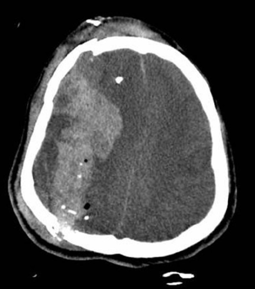
00:01 What about the evaluation of these patients? How do we diagnose and workup both the injury and potential secondary complications? Well, the first thing that should be done is a head CT. 00:12 Non-contrast head CT is the initial imaging modality of choice and we're looking for fractures, skull fractures, were looking for secondary injuries in the scalp, and intracranial hemorrhages. 00:24 Here we see a non-contrast head CT demonstrating a lens shape hyper density consistent with an epidural hematoma. 00:33 Alternatively, we may want to look in different planes and here we're looking at a coronal CT demonstrating a similar lens shaped deformity in the inferior temporal lobe also consistent with an epidural hematoma. 00:47 CTA or CT angiography is also important in evaluating these patients, particularly for concerned for dissection or tearing of the intima of the artery as a result of head or neck trauma. 01:00 Here we're looking at two CTA images. 01:02 The one on the left is a CTA in the sagittal plane, and we're looking at the contrast filling up the common carotids and internal and external carotids. 01:11 The common carotid here is labeled with the orange and we see the bifurcation of the internal and external carotid at the green arrow. 01:20 On the axial, we're looking at all four of the blood vessels that supply the brain with blood supply, the two common carotids in the green and the vertebral arteries in orange. 01:31 We can see the vertebral arteries are located within the foramen transversarium, the bones and that foramen of the bone in the cervical spine and that does increase the risk of tearing of that artery as a result of severe stress on the neck and selected injuries. 01:47 MRI scan is common to be performed in these patients. 01:50 And here we're not just looking for bleeding or fractures, but also internal injury to the brain itself. 01:57 This is an excellent modality for looking for a delayed onset of hemorrhage or other deficits within the first 24 to 48 hours after a trauma. 02:10 This is a modality that we may use for more moderate to severe concussions, but it's not required for all patients with a mild traumatic brain injury. 02:19 And here we're looking at a typical MRI scan, demonstrating a convex lesion extending over the entirety of the left convexity with multiple dense intensities of fluid consistent with an acute on subacute subdural hematoma. 02:36 This patient likely due to this left-sided brain hemorrhage will have right-sided weakness, maybe just drift with the arm or perhaps more extensive weakness, which could require surgical intervention. 02:50 We also want to consider monitoring for increased intracranial pressure as a result of trauma and swelling of the brain or hemorrhage. 02:59 We can see increases in intracranial pressure. 03:02 Intracranial bolt monitoring allows us to titrate intracranial pressure monitoring in the ICU setting for patients with significant or severe traumatic brain injuries. 03:12 And patients with a brain injury can have a seizure not all will. 03:16 But we evaluate for seizures clinically and in those who suffer a seizure treat promptly with an anti-epileptic. 03:24 So what about the initial management? How do we manage these patients? What are some of the initial interventions and then longer term interventions for patients? Well, the first is this brain injuries are an emergency and whenever we're dealing with an emergency, we think about airway protection. 03:39 Intubating those who may not be able to protect their airway, particularly those with a GCS scale of 9 or less. 03:47 Fluid resuscitation oxygenation are important. 03:50 We want to reduce biochemical stress to the brain. 03:53 And these resuscitative and supportive care measures are critical for doing that. 03:58 And then we want to assess and manage any associated injuries, injuries to the scalp, face, or the brain itself, or anywhere else in the body. 04:06 And patients presenting with a new trauma will have a head to toe evaluation to screen for any traumatic injuries anywhere in the body so that those can also be managed.
About the Lecture
The lecture Head Trauma: Diagnosis and Management by Roy Strowd, MD is from the course Head Trauma.
Included Quiz Questions
What is the initial imaging modality of choice in a patient with head trauma?
- Non-contrast CT of the head
- CT head with contrast
- CT angiography
- Non-contrast MRI of the head
- T2-weighted MRI of the head
What is the preferred imaging modality for a patient with a delayed diagnosis of head injury or severe concussion?
- MRI of the head
- Non-contrast head CT
- CT angiography
- PET scan
- EEG
What is the most important initial clinical consideration in patients presenting with new-onset head trauma?
- Intubation for patients with GCS less than or equal to 8
- Cervical spine X-ray
- Sedation to prevent seizures
- Mannitol to reduce intracranial pressure
- Blood transfusion
Author of lecture Head Trauma: Diagnosis and Management

Roy Strowd, MD
Customer reviews
5,0 of 5 stars
| 5 Stars |
|
5 |
| 4 Stars |
|
0 |
| 3 Stars |
|
0 |
| 2 Stars |
|
0 |
| 1 Star |
|
0 |
