Playlist
Show Playlist
Hide Playlist
Hard Palate

-
Slides Digestive system oral cavity.pdf
-
Reference List Histology.pdf
-
Download Lecture Overview
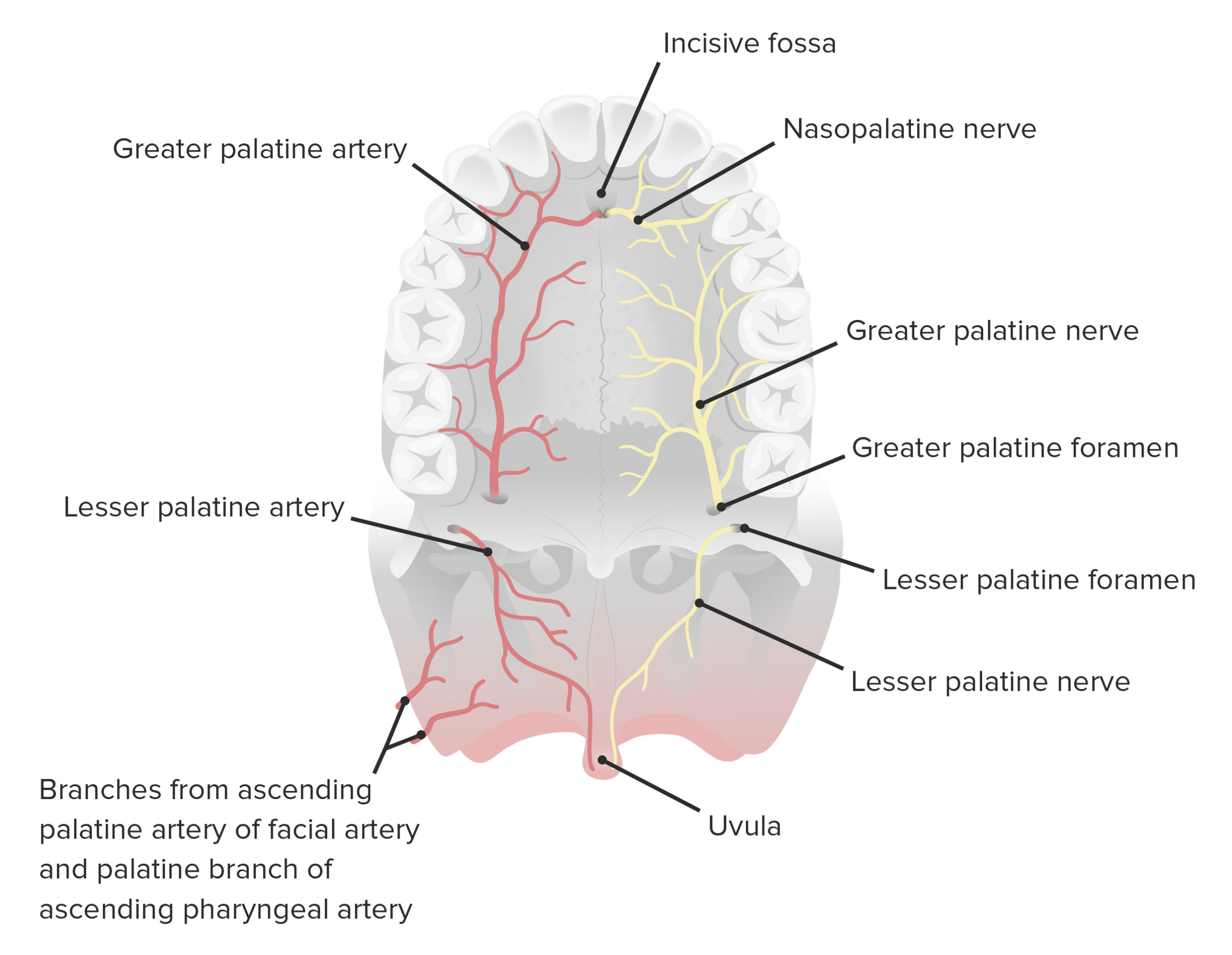
00:00 On this slide, you can see two sections taken through the hard palate. 00:06 On the left-hand side under very low magnification, you can see the hard palate labelled. It's actually bone. And on top of that bone, on the surface of the bone on the oral side is the masticatory mucosa that I'll describe in more detail in a moment. But superior to the bone of the hard palate, you can see two roughly even clearer structures. And in those clear areas, you can see two almost overlooking pale stained structures. This is the nasal cavity, and the two overlooking structures are in fact parts of the inferior concha. They are very important because they house respiratory tract epithelium, and also a very wide network of blood vessels called swell bodies that help warm, moisten and clean the air as it passes through the nasal cavity. And I've covered details of that in a lecture on the respiratory system. But focus now on the hard palate, and below, just below, you can see some rather pale stained structures on either side of the centre of the hard palate. They're mucus-secreting glands. A lot of glands secrete into the oral cavity to moisten it and also to help form that bolus of food I mentioned at the very beginning of the lecture. 01:40 On the far extreme of each end of this particular section are teeth. There's a tooth on the left section and a tooth on the right embedded in alveolar bone. And if you look very closely at this section, you can see a bright red component. That is the dentin of the tooth. And central to that dentin is a very pale region which is the pulp cavity. You don't see the enamel there because the enamel is a very mineralized structure. This section has been decalcified, and therefore, all you see here is the organic components remaining, and therefore, taking out the stain. Focus now on the right-hand section and on the masticatory mucosa. It has got a very thick epithelium, its stratified squamous epithelium. But if you look closely at this section, you might see a little bit of keratin on the surface. That can happen in the oral cavity where there is constant wear and tear, or constant abrasion. It's a bit above normal. For instance, if you wear a denture that doesn't fit properly, it can start to keratinize the epithelial surface at that location. We call it para keratinized because it doesn't go through the full keratinization process. But generally speaking, the oral mucosa and the masticatory mucosa on the surface of the hard palate is just stratified squamous epithelium. 03:27 Notice the epithelium actually embeds into the underlying connective tissue, and the underlying connective tissue is very dense collagen. So this epithelial surface is very tightly bound to the bone. 03:41 It forms a very, almost hard surface for the tongue to push food up against and help to mechanically break it down.
About the Lecture
The lecture Hard Palate by Geoffrey Meyer, PhD is from the course Gastrointestinal Histology.
Included Quiz Questions
Where does the nasal cavity lie?
- Superior to the hard palate
- Superior to the cribriform plate
- Posterior to the cribriform plate
- Posterior to the hard plate
- Inferior to the soft palate
Which of the following is the normally visible part of the tooth?
- Enamel covering the crown
- Root
- Pulp
- Dentin
- Vascular supply of the tooth
Which of the following describes the epithelium of the masticatory mucosa?
- Stratified squamous epithelium
- Simple columnar epithelium
- Nonkeratinized simple squamous epithelium
- Stratified columnar epithelium
- Simple cuboidal epithelium
Which ONE of the following is INCORRECT?
- The masticatory mucosa lines the inside of the cheeks and the floor of the mouth.
- Teeth are embedded in alveolar bone.
- Lining mucosa is nonkeratinized stratified squamous epithelium.
- Venous plexuses in nasal cavities warm the inspired air.
- Gland secretions into the oral cavity assist in the formation of food boluses.
Author of lecture Hard Palate

Geoffrey Meyer, PhD
Customer reviews
5,0 of 5 stars
| 5 Stars |
|
5 |
| 4 Stars |
|
0 |
| 3 Stars |
|
0 |
| 2 Stars |
|
0 |
| 1 Star |
|
0 |
