Playlist
Show Playlist
Hide Playlist
Gardnerella Vaginalis

-
01-20 Gardnerella Vaginalis.pdf
-
Download Lecture Overview
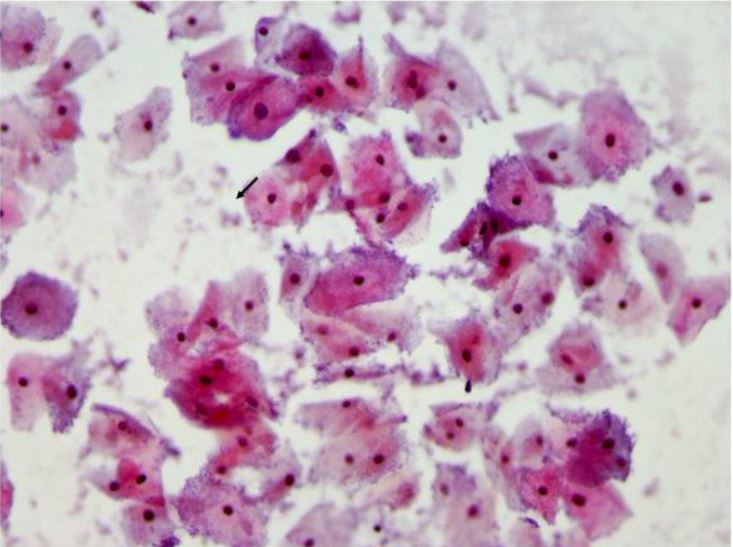
00:01 Gardnerella Vaginalis; a bacteria. Gardnerella are small pleomorphic gram-variable bacilli. 00:09 Pleomorphic because they can appear not just as caucus but sometimes as a more oval-shaped. 00:17 And gram-variable because the gram up take - gram stain uptake may be a dark purple as a gram-positive or a pink as a gram-negative and they can appear sometimes as both, sometimes in the same organism. 00:31 The image on the slide projects a single Gardnerella organism which in this instance looks quite caucus or caucal in shape; rounded. 00:40 But if one were to look at it by switching the image just 90°, it might appear much more elongated or rodlike. 00:49 Regardless, Gardnerella are none spore forming and they're nonmotile; they stay where they're placed when they grow. 00:56 They are facultatively anaerobic and they are one of the causes of bacterial vaginosis. 01:03 That particular entity frequently is polymicrobial caused by many different bacteria. 01:08 But as we'll see; the Gardnerella are fairly prominent component of the diagnosis of a bacterial vaginosis. 01:17 How does the pathogenesis occur? Typically, the organism will adhere to its receptor sites, penetrating through the outer layers of vaginal mucosa, via vaginolysin. 01:29 Once attached to underlying epithelium, the Gardnerella bacteria are able to multiply and then hyper secrete or form a biofilm; its own sort of proteinaceous and polysaccharide rich covering which further prevents it from immunologic discovery and immune response. 01:49 While there under the biofilm, the Gardnerella compete with normal vaginal flora such as Lactobacillus to dominate the overall vaginal flora. 02:00 Once that occurs, then escalation can occur in the vaginitis. 02:05 What are the clinical manifestations? Typically, as the Gardnerella emerges and starts to hyper produce and hyper secrete, a nonpainful, but a grayish vaginal discharge will develop. 02:18 And here's the icky part and the prominent part. It smells like dead fish. 02:23 Yes, Gardnerella is known for having a fishy smell and its presence while distinguished from Trichomonas which also can create that fishy smell is certainly a suggestion that a bacterial vaginitis or vaginalis is ongoing. 02:40 The images you see on the screen simply show a multiplication of the Gardnerella. 02:44 And here as multiplying, they appear more elongated, more in a rod shaped form. 02:51 How do we make the diagnosis? Well, we're fortunate, in being Sherlock Holmes that we have clue cells. 02:59 A clue sell is a vaginal epithelial cell which is coded by attached or bound Gardnerella bacteria such as you see in the image on the screen. 03:10 You can distinguish the clue cell, so noted with the arrow from other vaginal epithelial cells on the right side of the image which have only a few bacteria attached to them. 03:22 In addition, the amine whiff test is quite significant. 03:27 In this, one mixes that gray vaginal discharge with 10% potassium hydroxide and that will further enhance the fishy smell just in case anybody had any doubts about its presence in the first place. 03:40 Treatment for Gardnerella involves typically the antibiotic metronidazole as a drug with excellent anaerobic coverage. 03:48 Although, clindamycin also can be used as this continues the anaerobic coverage significantly. 03:56 So Gardnerella, the organism with a fishy smell and something to be quite aware of although distinguished from Trichomonas, but it contributes significantly to bacterial vaginitis.
About the Lecture
The lecture Gardnerella Vaginalis by Sean Elliott, MD is from the course Bacteria.
Included Quiz Questions
What are the features of vaginal discharge which is caused by Gardnerella vaginalis infection?
- Nonpainful, gray discharge with fishy smell
- Painful, gray discharge with fishy smell
- Painful, yellow discharge with fishy smell
- Nonpainful, gray discharge with pungent smell
- Nonpainful, yellow discharge with pungent smell
Diagnosis of Gardnerella vaginalis infection can be made by the presence of which type of cells seen with a microscope?
- Clue cells
- Foam cells
- Kulchitsky cells
- Multinucleated giant cells
- Oxyntic cells
Author of lecture Gardnerella Vaginalis

Sean Elliott, MD
Customer reviews
5,0 of 5 stars
| 5 Stars |
|
5 |
| 4 Stars |
|
0 |
| 3 Stars |
|
0 |
| 2 Stars |
|
0 |
| 1 Star |
|
0 |
