Playlist
Show Playlist
Hide Playlist
First-time Seizure: Evaluation, History, and Examination

-
Slides Seizures Epilepsy New Onset Seizure.pdf
-
Download Lecture Overview
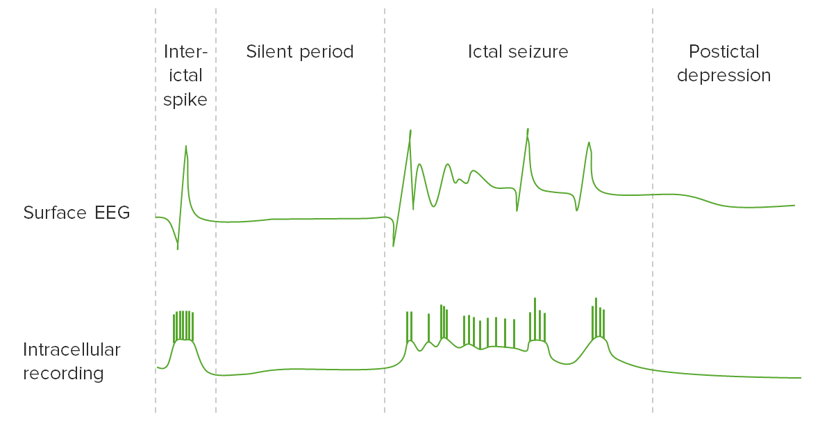
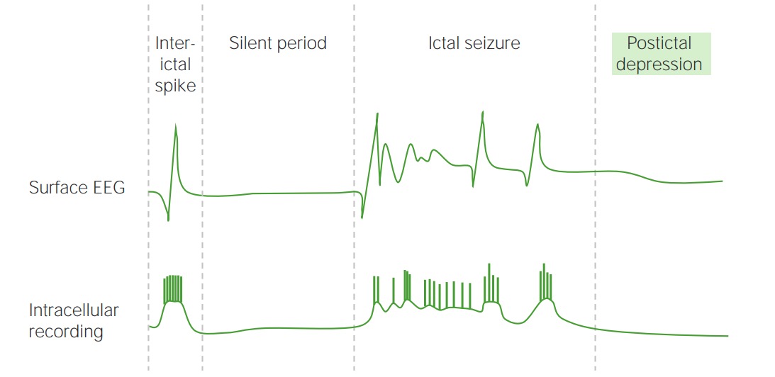
00:01 So how do we evaluate the first time seizure? Starts with a history and we need a good seizure semiology to understand the type of seizure, we look at the patient's prior history and family history. 00:13 Following a history and physical examination will conduct laboratory evaluation just like any other patient. 00:18 Neuroimaging is key, and nearly all patients who were evaluating for seizures will undergo an EEG or electroencephalography. 00:27 The goal of evaluating a first time seizure is to figure out the risk of a second seizure. 00:33 So William Gowers, one of the famous neurologist said, seizures beget seizures, and he's right, the more seizure a patient suffer, the more likely they are to suffer long term seizures. 00:43 So we want to reduce the risk of a second seizure. 00:47 And the tests we do on physical examination in history, as well as the diagnostic investigations with imaging and EG are really to differentiate the patient's risk of developing a second seizure. 00:58 In general, the risk of recurrent seizure after a first unprovoked seizure is 40%. 01:04 40% of patients will have a second seizure, which is low, and this is why we don't treat all first time seizures. 01:11 The risk of a recurrent seizure after a second unprovoked seizure is upwards of 75%. 01:18 If you've had two seizures, the likelihood of having a third is 75%. 01:22 If you've had three, that number is more than 80 or 90%. 01:25 And so we treat after the second seizure. 01:28 The goal of evaluation is to stratify the risk of recurrence. 01:32 Is this patient with a first time seizure likely to suffer a second time seizure. 01:37 If that risk is greater than 50%, we may treat. 01:40 If it's less than 50%, as with the standard, healthy adult, we would not. 01:45 And the goal of treatment is to prevent and manage recurrent events, prevent the risk of future seizures. 01:53 So let's talk through the history and examination and understand what we do to evaluate the first time seizure. 01:58 On history, we're looking at the aura, the pre-ictal process, vocal motor, respiratory and autonomic signs that may point us to the seizure semiology, we look for precipitants, particularly if they're treatable electrolyte abnormalities, fever and infectious symptoms or new medications. 02:16 We ask about and look for ictal automatisms, common automatisms these are automatic behaviors that occur during the seizure and can tip us off to whether other seizures may have occurred are chewing, swallowing, eating, gestures, ambulation and mimicry all of those we can see. 02:33 There are also some automatisms that are associated with absence seizures and may include scratching or fumbling with clothes or lingual movements and other complex motor activities. 02:45 On examination, we're looking for neurologic deficits or focal abnormalities that indicate the location of the seizure, the lateralization of the seizure or potential co-occurring phenomenon, a stroke in addition to seizure. 02:58 When we ask about and look at ictal symptoms, we're trying to figure out, what is the lateralization? What's the side of the brain that's affected, that helps us to approach imaging and for patients that fail medications and we're considering surgery will point us to the side to consider. 03:13 We're also looking for things that will help us to localize. 03:16 This is more advanced in lateralization, not just which side of the brain is the seizure coming from, but what's the precise focus? Can we get it into a specific lobe of the brain? Can we hone in even further on a specific area or cortex area of cortex of the brain? And some of the clinical descriptions can help hone us in on those. 03:37 Head turning which is called head version can be a helpful lateralizing and localizing sign. 03:43 Early non-forced head version is typically seen from an ipsilateral temporal origin, forced early head version from ipsilateral frontal origin, late forced head versions so that's a head moving to one side, late in the seizure is seen with contralateral frontal or temporal origin. 04:00 And you can see a number of other findings, ocular version that's moving of the eyes to one side, focal clonic activity or dystonic limb, unilateral tonic limb, a M2e to sign which is a fencing posture can be seen with frontal, contralateral frontal seizure origin, a figure 4 shape of the hands can be seen again with contralateral hemisphere seizure onset. 04:24 Ictal paresis is seen in the contralateral motor cortex. 04:29 Todd's paralysis is common after focal onset seizures involving the motor cortex and points us in the direction of the contralateral motor cortex. 04:37 Unilateral blinking, unilateral limb automatisms from the ipsilateral hemisphere. 04:43 Postictal nose rubbing is a localizing finding, localizing to the ipsilateral temporal lobe postictal cough, bipedal abnormalities, hypermotor activity, ictal spitting can be lateralizing to the right side, as well as the automatism with reserved responsiveness in the right temporal lobe. 05:04 So we use these features to figure out which side of the brain and which location in the brain the seizures may be coming from, which can help us in orienteering evaluation and subsequently treatment. 05:15 And I've included here some other localizing collateralizing sign, gelastic seizures with laugh can be seen in hypothalamic areas of onset. 05:25 And you can see here it does urge and speech arrest and post ictal aphasia. 05:31 So if that's how the seizure begins, we also want to interrogate what happens during the event, the semiology of the event, and we think about other signs that can help us to understand where the seizure may be coming from during the event. 05:43 Ictal vomiting and the insular cortex paraesthesia from the sensory cortex, clonic or tonic clonic activity involving the motor cortex. 05:51 Dystonic posturing points to a frontal lobe seizure, 60% of auras involve sensory symptoms, the parietal cortex is commonly affected. 06:00 89% of patients with frontal lobe epilepsy will have dystonic posturing, sometimes with retained awareness out of sleep which can be quite jarring to patients and or other caregivers. 06:12 And then early head version we said localizes and late of the head version lateralized. 06:18 A fencing posturing can be seen from seizures originated in the supplementary motor area. 06:23 Again, early head version is ipsilateral origin, late head version is from a contralateral origin. 06:29 And pain can be seen from a contralateral sensory cortex seizure. 06:35 Olfactory symptoms or automatisms, or olfactory smells can be seen with seizures originating from the orbital frontal cortex. 06:42 Ictal spit, piloerection localized to the temporal lobe, lateral temporal lobe, we can see Deja vu early in the seizure as the aura and with medial temporal lobe, abdominal fear a rising sense of nausea patients can describe.
About the Lecture
The lecture First-time Seizure: Evaluation, History, and Examination by Roy Strowd, MD is from the course Seizures and Epilepsy.
Included Quiz Questions
Which statement regarding new-onset seizures is most accurate?
- The primary goal of evaluating the first episode is to assess a patient's risk for subsequent seizures.
- Patients usually begin treatment after the first unprovoked seizure.
- After the first episode, the risk of a second seizure is around 75%.
- Localization is rarely attempted as it offers little therapeutic value.
- After a patient experiences a second seizure, there is little risk of suffering a third seizure.
Which statement is the most accurate in terms of localization?
- Localization plays a role in the therapeutic options available to a patient.
- Localization of a seizure depends entirely on the neurologic exam.
- Lateralization information is primarily for research purposes.
- Only seizures originating from the temporal lobe can be localized.
- History of symptoms from a prior episode is of little clinical value.
Which statement regarding seizure symptoms is the most accurate?
- Laughing may indicate hypothalamus involvement.
- Early head-turning suggests lateralization.
- Ictal splitting localizes to the left temporal lobe.
- Piloerection indicates frontal lobe involvement.
- A postictal cough suggests frontal lobe involvement.
Author of lecture First-time Seizure: Evaluation, History, and Examination

Roy Strowd, MD
Customer reviews
5,0 of 5 stars
| 5 Stars |
|
5 |
| 4 Stars |
|
0 |
| 3 Stars |
|
0 |
| 2 Stars |
|
0 |
| 1 Star |
|
0 |
