Playlist
Show Playlist
Hide Playlist
Femoral Triangle – Anterior and Medial Thigh
-
Slides 05 LowerLimbAnatomy Pickering.pdf
-
Download Lecture Overview
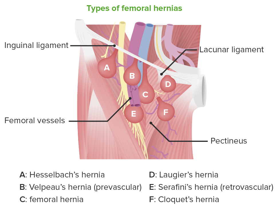
00:01 Now, I want to talk about the femoral triangle. 00:04 This is an important region that contains primarily the femoral nerve, the femoral artery, and the femoral vein. We can see that it’s a triangle and it’s positioned approximately here. We’ve got borders from the adductor longus muscle, from the sartorius muscle, and from the inguinal ligament. The roof of it is covered by the fascia lata. And importantly, we have the saphenous opening which allows the great saphenous vein to pass in. 00:35 So the femoral triangle is an important subfascial space located in the upper thigh. The boundaries of it, we have superiorly and we have the base of this triangle, and that’s going to be the inguinal ligament, which we can see here. We then taper down into this apex. 00:55 Laterally, we have sartorius. So the lateral boundary of the femoral triangle is the medial border of sartorius. The medial boundary of the femoral triangle is going to be the lateral border of adductor longus. So here we have this medial boundary, we have this lateral boundary, and we have this superior boundary. And that forms this triangle that is known as the femoral triangle. The floor of the femoral triangle is formed by two muscles - iliopsoas and pectineus. Laterally, we find iliopsoas. Medially, we find pectineus. 01:37 And these are the boundaries and the floor of the femoral triangle. Within the femoral triangle, we have a number of structures. We have the femoral nerve, the femoral artery, and the femoral vein. Importantly, the roof is formed by the fascia lata. And here, we can see the femoral vein, and we see an opening in the fascia lata here. And this fascia lata, this opening is known as the saphenous opening, and this is how the medially positioned great saphenous vein can pass into the femoral vein. So we got this opening here. We can see the contents of the femoral triangle include from lateral to medial. We have the femoral nerve. 02:23 We have the femoral artery. We have the femoral vein. And then not included here, we have the deep inguinal lymph nodes. So we have nerve, artery, vein, inguinal lymph nodes from lateral to medial within the femoral triangle. This femoral triangle receives the femoral artery and the vein and the femoral nerve, which are passed from the abdomen, and they enter into the lower limb via the retro-inguinal space. The retro-inguinal space is a passage that connects the trunk with the lower limb. We can see that the retro-inguinal space lies behind the inguinal ligament. So we can see the inguinal ligament here. 03:09 It’s running from the anterior superior iliac spine all the way down to the pubic tubercle. 03:16 And we can see it divides the retro-inguinal space or the retro-inguinal space is divided into two compartments - a lateral compartment which we can see here, and that contains iliopsoas and the femoral nerve passing through. So we can see this iliopsoas and the femoral nerve passing through. Here, separating the two compartments is the iliopectineal arch, and we can see that running down separating this lateral compartment into this medial compartment. The medial compartment has the femoral artery, the femoral vein, and the lymphatics passing through, and we can see those here. And this is all occupied within the retro-inguinal space. It’s how these structures pass from the trunk into the lower limb.
About the Lecture
The lecture Femoral Triangle – Anterior and Medial Thigh by James Pickering, PhD is from the course Lower Limb Anatomy [Archive].
Included Quiz Questions
Which structure forms the medial boundary of the femoral triangle?
- Adductor longus muscle
- Fascia lata
- Saphenous vein
- Inguinal ligament
- Sartorius
What is the boundary of the sartorius muscle within the femoral triangle?
- Lateral
- Medial
- Floor
- Superficial
- Deep
Which of the following is contained within the femoral triangle?
- Femoral nerve
- Celiac trunk
- Cauda equina
- External iliac vein
- Popliteal artery
Customer reviews
5,0 of 5 stars
| 5 Stars |
|
5 |
| 4 Stars |
|
0 |
| 3 Stars |
|
0 |
| 2 Stars |
|
0 |
| 1 Star |
|
0 |


