Playlist
Show Playlist
Hide Playlist
Examination of the Cranial Nerves VII – XII

-
Reference List Physical Examination.pdf
-
Download Lecture Overview
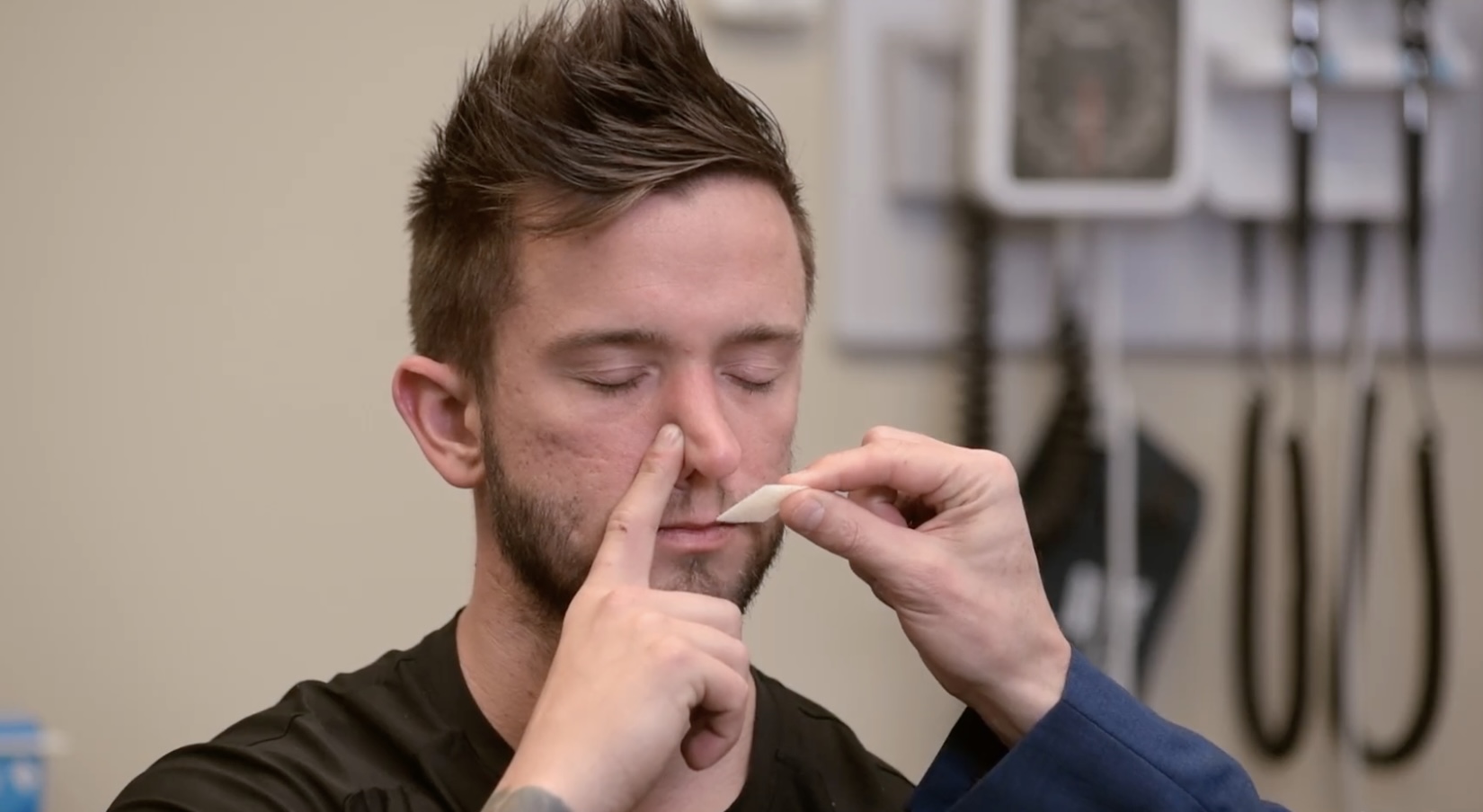
00:00 Alright, so having completed the 5th cranial nerve, we already talked about the 6th cranial nerve, so we're now moving on to the 7th cranial nerve, the facial nerve. The facial nerve has motor, sensory, and even autonomic nervous system function, but the only parts that we can really assess on physical exam is the facial function. So let's jump into that. The facial nerve when it exits just anterior to the ear and goes to the parotid gland, there's a number of different branches working at different muscles in the face. Even before it exited anterior to the ear, it went off and innervated the stapedius muscle within the middle ear. "So to test the facial nerve, I'm going to have you first lift up your eyebrows. Great. That's testing the frontalis muscle. Now vigorously lift up your eyebrows. Great. Now close your eyes really tightly. This is the orbicularis oculi muscles." Then I'm going to have him puff out his cheeks. 00:52 "Great. And don't let me push them. Perfect. And now have you smile. Great." So, this is a quick way to test for the different innervations of the facial nerve to the muscles of the face. 01:04 Importantly, patients who have a peripheral lesion with a facial nerve palsy, the classic one being Bell's palsy, will have weakness to the entire half of their face. In contrast, a patient who has had a stroke, it turns out that the frontalis muscle is duly innervated by both hemispheres. So, if you had a stroke on the left, yes you'll have weakness on the left side of your face if the stroke was in your right cerebral cortex, but because the left cerebral cortex also innervates the frontalis muscle you would still have intact movement of the frontalis muscle. So that can be a telltale sign to identify a central lesion versus a peripheral lesion and of course those are very different approaches that you'll take based on which of those you've identified. Next up, we'll move off and do the vestibular cochlear nerve, that is the 8th cranial nerve. We're going to start off by assessing the hearing part of the vestibular cochlear nerve, that's the cochlear branch and then we'll come to the vestibular nerve afterwards. So, to assess hearing, let's start off by just gross hearing and that's simply just "Can you hear this on both sides?" Just the rubbing of the fingers together can just elicit a soft sound that patients with grossly intact hearing should be able to discern. If there are any concerns when you perform that test or if the patient reports or having difficulty hearing, then it's time to perform the Weber and Rinne tests. Ideally you want to use a 512. 02:31 You could also use a 256. Patients who are older oftentimes will lose the ability to hear higher pitches like a 512, and that's sort of a normal part of aging. It's presbycusis that loss of those very high pitches. Both of these tests are designed to distinguish between conductive hearing loss versus sensorineural hearing loss. And conductive hearing loss would be if he even had a lot of wax in his ears that was occluding the external acoustic meatus, then the sound that I make out here is simply not going to get to the eardrum. Or if there is a problem with the perforated eardrum or if there is a problem with the ossicles of the bones that are magnifying the sound and transferring it or passing it on to the sensory nerve, to the cochlear nerve. So, conductive hearing loss versus sensorineural hearing loss, that's where we use these tests for. I always remember which one of these tests is the Weber, the Weber test is going to be up here. Weber starts with a W and that's a symmetric letter and so I can remember I'm starting in the middle of his forehead and we'll do that now by making a loud sound. "Which side do you hear that the most on or is it pretty much the same?" "A little harder." "Sure." "Both." "Great." So, if he had a problem with conductive hearing loss, again let's say that his left ear was completely full of cerumen and he was unable to get any sound moving to his sensory nerve, his cochlear nerve. His cochlear nerve on the left would be starving for information and so would amplify its sensitivity to sound and so when I put this on his forehead he would lateralize to the left if he had a conductive hearing problem. 04:14 He would try and his left cochlea would be amplifying its detection of sound even though it's vibrating through his scalp. In contrast, if he had a sensorineural problem let's say on the left where the nerve was out perhaps due to a, like a schwannoma or a viral labyrinthitis, something that's causing damage to the nerve itself that when I want to put this on his forehead he would lateralize to the right because the left sensory input is not functioning, the detector is not functioning. So this test doesn't tell us what the cause of the problem is because you need to do the next step in this test and that's the Rinne exam. The Rinne exam is performed as follows. "Can you hear that?" "Yes." "Let me know when you stop hearing it." "Stop." "Can you hear it now?" "Yes." "Great." So, what we've done there is we've used his mastoid process just where I put the head of this tuning fork to test for bone conduction. 05:31 Bone conduction is essentially seeing if the sensory nerve is still able to gather information whereas when I hold the tuning fork out here, this is air conduction which relies upon, again, the machinery of hearing. If under normal circumstances air conduction should be better than bone conduction because in general the ears were designed to magnify information coming from sound and that's what the ossicles and the tympanic membrane are designed to do as opposed to getting sound just from vibrations on your skull, which is what I'm testing with bone conduction. So again, if his ear is full of cerumen, he's going to have decreased air conduction because the sound of vibrations from the air is just not going to penetrate pass there so when I did this test and waited till he could no longer hear via bone conduction when I would bring the tuning fork out here he would not have been able to hear it and then I would know especially if the Weber test had localized to the left side that his problem is a conductive hearing loss problem. So, those are the 2 tests that we use to really dive in and identify conductive versus sensorineural hearing loss. Now we're going to assess the vestibular portion of the vestibular cochlear nerve and those 2 tests that we're going to highlight here. One is the head impulse test and we're going to do the Dix-Hallpike test now. 06:55 Patients with benign paroxysmal positional vertigo will commonly have to undergo this test to sort out exactly what's causing their symptoms and in general the idea is that there is a little otolith, a little stone folding around in the semicircular canals that's causing these paroxysms of vertigo. By doing this maneuver that I'm going to demonstrate, we're trying to have one of those otoliths quickly dislodged and moved around in the semicircular canals to reproduce the symptoms and potentially the nystagmus that can come along with BPPV. "So, I'm going to start off by having you sit up for me please. And scoop back just 2-3 inches or so. Great. And what I'm going to have you do is look towards your left and I'm going to lower you down quickly and we're going to have your head a little bit off the table. Ready, go. 07:45 Good." Now what I'm looking for here is I'm going to have him look off into the distance and we'll keep him here for about 30 seconds or so and we're looking for either the reproduction of symptoms. So his subjective report of having either nausea or vertiginous symptoms and I'm also looking for nystagmus. The Dix-Hallpike maneuver is designed to try and elicit nystagmus. It can take up to 30 seconds to 60 seconds to actually bring it out, but it would be very supportive of a peripheral cause of vertigo namely BPPV. "Okay, we'll have you sit up." I would do that on both sides, but now what we'll do is the head impulse test. "And you can face forward now." Alright, so the next test that we're going to do to assess the vestibular nerve is called the head impulse test. And this is tricky because you don't want to perform this test on a person who doesn't have any symptoms, any symptoms of vertigo, etc. because it's going to be normal in a person who does not have symptoms but it will also be normal in a person who has had a stroke. So, you're really using this test just to identify in a person who has symptoms whether they're having a stroke or they just have a problem with the vestibular nerve itself typically a vestibular neuritis or something benign that's going to resolve within a matter of hours to days. So, with that said let me demonstrate what the head impulse test is and then we will interpret it. "So let me have you look over at my nose and what I'm going to do is just gently move your head around in space just to get your neck nice and pliable. Then I'm going to quickly move your head in 1 direction. Good." And I want you to take note that his oculocephalic reflex is intact no matter how quickly I move him. His eyes stay locked on to my nose. And if his eyes are staying locked on to my nose, I know that his vestibular nerve is functioning. In contrast, if a patient is having vertigo symptoms with or without nystagmus, they're going to misdirect, their eyes are going to follow the direction of the head and they'll have to make a corrective saccade back to my nose. In contrast, a person who is having a stroke, they have dual inputs, they have both sides of their head are working to make sure the oculocephalic reflex is working and if the vestibular nerves are intact they will also have a normal finding. So it's important to make sure you're just doing this test in someone who is actively having vertiginous symptoms. Alright, with that we can move on to cranial nerves IX and X. So cranial nerve IX is the glossopharyngeal nerve, cranial nerve X is the vagus nerve, and we're going to do them together. The vagus nerve has a lot of other things that it does in terms of the autonomic nervous system, but for the purposes of the bedside physical exam we're really focusing on its role in terms of regulating swallowing. So, these different nerves are difficult to distinguish their actions when we're looking at the swallow reflex but they are going to help us, we can identify that they're both out or not using these maneuvers. So let me first grab my penlight, my otoscope light. So the glossopharyngeal and vagus nerves are going to help with elevation of the soft palate. So let's take a look. "Say ah." "Ahhh." "Great." We can see that there is symmetry between the arches and the back. There is soft palate is going up. We also see that his uvula is also midline. In a patient for whom there was evidence of asymmetry, one side is not going up the way that it should. We could then test the gag reflex which we can either do with a Q-tip swab and just touch the back of the throat or tickle the uvula to make sure that the gag reflex is intact. Alright, we're on the home stretch with the cranial nerve exam. We're on cranial nerve XI. This is the accessory nerve and this nerve innervates 2 important muscles and it's the sternocleidomastoids and the trapezius muscles. 11:38 "To test those, I'm just going to have you turn your head to the left against resistance. 11:43 Great." And keep in mind that him turning his head to the left is actually the function of his right sternocleidomastoid muscle which you could see was tensing up there. "I'm going to have you turn your head to the right. Great." And that's his left sternocleidomastoid muscle shown there and then the muscles of the trapezius are operating to elevate his head or laterally flex his head as well depending upon the orientation of his head at the time. So, next up is the XII cranial nerve, the last one, this is the hypoglossal nerve and it's simply tested by having the patient "stick your tongue out please and move it from side to side." "You could put your tongue back." A patient who has a defect in the XII cranial nerve on the left will actually deviate their tongue towards the affected side because that nerve is designed to make the tongue muscle push out. So if it's not working, it's going to end up being dominated by the right hypoglossal nerve and the musculature once you push the tongue off to the left.
About the Lecture
The lecture Examination of the Cranial Nerves VII – XII by Stephen Holt, MD, MS is from the course Examination of the Nervous System.
Included Quiz Questions
Which cranial nerve (CN) do the Weber and Rinne tests evaluate?
- CN VIII: cochlear nerve
- CN IX: glossopharyngeal nerve
- CN X: vagus nerve
- CN XI: accessory nerve
- CN XII: hypoglossal nerve
Which cranial nerves (CN) are tested by asking the patient to elevate their palate by saying “AH?"
- CN X: vagus nerve and CN IX: glossopharyngeal nerve
- CN XI: accessory nerve and CN XII: hypoglossal
- CN V: trigeminal nerve and CN VII: facial nerve
- CN VIII: cochlear nerve and CN VII: facial nerve
- CN III: occulomotor nerve and CN V: trigeminal nerve
Which cranial nerve (CN) is tested by asking the patient to turn their head against resistance?
- CN XI: accessory nerve
- CN IX: glossopharyngeal nerve
- CN VII: facial nerve
- CN X: vagus nerve
- CN VIII: cochlear nerve
Which cranial nerve (CN) is tested by asking the patient to move their tongue from side to side?
- CN XII: hypoglossal nerve
- CN X: vagus nerve
- CN XI: accessory nerve
- CN VII: facial nerve
- CN IX: glossopharyngeal nerve
Which cranial nerve (CN) is affected by Bell’s palsy?
- CN VII: facial nerve
- CN IX: glossopharyngeal nerve
- CN XI: accessory nerve
- CN XII: hypoglossal nerve
- CN X: vagus nerve
Which cranial nerve (CN) provides sensory innervation to the palate and is tested using the gag reflex?
- CN IX: glossopharyngeal nerve
- CN XI: accessory nerve
- CN XII: hypoglossal nerve
- CN VII: facial nerve
- CN VIII: cochlear nerve
Author of lecture Examination of the Cranial Nerves VII – XII

Stephen Holt, MD, MS
Customer reviews
5,0 of 5 stars
| 5 Stars |
|
1 |
| 4 Stars |
|
0 |
| 3 Stars |
|
0 |
| 2 Stars |
|
0 |
| 1 Star |
|
0 |
Great lesson, a very useful summary for everyday clinical practicing.
