Playlist
Show Playlist
Hide Playlist
Etiologies of Neck and Back Pain
-
Slides Physical Exam Neck and Back Pain Introduction.pdf
-
Reference List Physical Examination.pdf
-
Download Lecture Overview
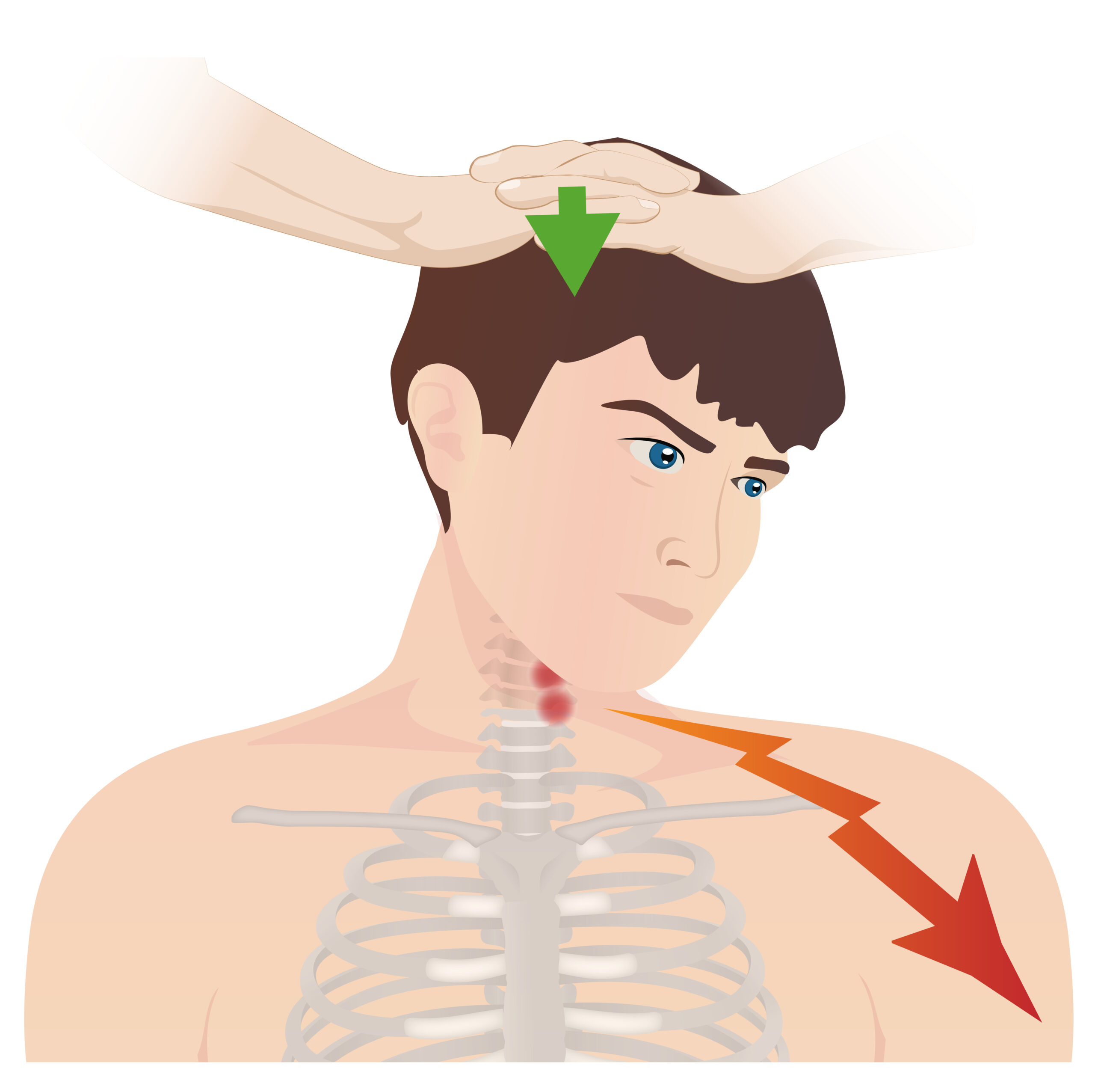
00:01 So here's an important way to frame the next few slides. 00:05 In the absence of nerve root impingement or spinal cord compression, is all atraumatic neck pain necessarily due to muscle or tendon strain? The answer is no, this is false. 00:20 If you will look at this X-ray of this knee, I'd think you'd see that there's a lot of disease in this X-ray, for example, you could see that there's loss of the joint space and the lateral compartment. 00:34 There's sporadic changes happening to the bone there and there's subchondral cysts. 00:38 And there'd be no surprise that a person with this degree of knee osteoarthritis, every step is causing them pain. 00:44 But there's no nerve roots passing between those bones, so why does this person have knee pain? Remember that every structure in the back is innervated, every bone, so while oftentimes we're thinking that this person who has low back pain can either have a muscular problem or a nerve root that's being pinched, remember that all those facets, the ligamentum flavum, the posterior longitudinal ligament, every structure is innervated and can cause low back pain. 01:15 It's important for us as the examiners to distinguish, is this just pain limited to those structures or is there actually disease involving the spinal cord or the nerve roots because that's going to change our management. 01:27 Shown here in this picture, particularly one in the middle is spinal stenosis and this is the end stage of spondylosis where there's so much disease in the spinal canal that's encroaching and actually causing edema of the spinal cord itself. 01:40 This is what we're looking for in patients with back pain and we want to exclude it from our physical exam. 01:46 In contrast, a lot of people who have pain in their back, simply have arthritis of these facets shown here, so-called facet joint arthropathy or degenerative disease of the facets. 02:00 Another example of spondylosis is what's shown here on the right of the figure which is spondylolisthesis, which can be either on the micro level or the macro level, and it's one vertebral body sliding over another, and you could imagine how if its significant spondylolisthesis that can pinch the spinal cord at that level as well. 02:19 When patients just have facet joint arthropathy or disease involving the bones, people will have axial pain that is just referred pain radiating to the back itself, whether it's cervical areas shown on the left or the thoracic areas shown on the right, and then further down would be the lumbar areas, that's where the pain is going to be localized to. 02:41 So, this slide shows the different ways that radiculopathy can occur. 02:47 So first off on the left-hand side of this figure, we see what's called an uncovertebral join osteophyte. 02:53 I talked about facet joints, there's also uncovertebral joints which is basically the articulation between one vertebral body and the next. 03:02 If there's an osteophyte or again a bony outgrowth which accompanies osteoarthritis, in that joint, it will encroach upon the nerve root that is exiting through the right-sided neural foramina there, that's going to cause a radiculopathy downstream. 03:20 Likewise, if the nucleus pulposus, the white matter inside of an intervertebral disk herniates as demonstrated here also on the left side of this image, it also can encroach upon that nerve root and cause downstream radiculopathy. 03:34 And lastly, on the right side of the figure, you can see a facet joint osteophyte that is also pinching and encroaching upon those nerve roots as they exit. 03:44 You can imagine that the nucleus pulposus could also herniate more into the central area of the canal and encroach upon the spinal cord itself. 03:54 So, as we'll see more in our physical exam assessment, cervical radiculopathy is going to be exacerbated by any maneuver that increases tension on the nerve roots and also anything that will further narrow the neural foramina itself. 04:11 The most commonly affected nerve roots are the C6, C7, and the C5 nerve roots and we're going to have to, in our physical exam, figure out how to identify problems with those specific nerve roots. 04:23 Moving on to the lumbar radiculopathy assessment. 04:27 Again, it's going to be exacerbated by any maneuvers that can increase tension on the nerve roots, anything that further narrows the neural foramina, and the nerve roots that are most commonly affected for lumbar radiculopathy are the S1 and L5 nerve roots; but L4, L3, and even L2 can also be affected with radiculopathy. 04:48 I've already alluded to the possibility that the spinal cord itself can be affected with any type of spondylosis but in particular, spondylolisthesis, where one vertebrate is sliding over the other. 05:03 And we're going to look for physical exam findings that support involvement of the spinal cord. 05:08 Patients with spinal cord involvement will oftentimes have ataxia, clumsiness in their upper extremities. 05:14 We're looking for a combination of upper motor neuron and lower motor neuron manifestations. 05:20 Typically with spinal stenosis, there's multiple nerve roots involved, those at the level of the injury and also below; and you'll also have atrophy and leg weakness in most patients who have advanced spinal stenosis. 05:35 So while we've been focusing on some of the more indolent progressive disease processes of the spine like spinal stenosis, you can also, of course, have patients presenting with acute trauma for example after a motor vehicle accident. 05:50 Some of the more common places where a fracture of a cervical spine vertebrate can occur is the dens of the atlas. 05:56 You can also have compression fractures which can be from osteoporosis which is so-called insufficiency fractures or could also occur in a setting of trauma, and patients may have exquisite tenderness over particular vertebrate as you marched down the posterior aspect of the spine. 06:13 Likewise there could be structural defects which may be congenital, scoliosis. 06:18 We're going to look for particular exam findings and how to bring that about on the exam. 06:22 Kyphosis, the natural curvature of the spine. 06:26 Lordosis, similarly can be exaggerated in certain circumstances. 06:30 And lastly, as I alluded to before, there are absolutely other systemic diseases that may manifest in the spine, in particular, rheumatoid arthritis, more commonly involves the C spine and then ankylosing spondylitis which is going to be more commonly affecting the sacroiliac joints and the lumbar spine. 06:53 We'll also, in the next lecture, look at the hip and sacroiliac joints which, in patients with low back pain, is a required part of our assessment. 07:04 Patients may have anterior etiologies for their hip pain listed below. 07:11 Lateral causes of hip pain and then particularly, when we're thinking about patients with low back pain, we want to assess for these posterior etiologies such as sacroiliitis, piriformis syndrome and ischial tuberosity bursitis. 07:26 A quick review of the anatomy of the hips and the SI joints. 07:31 It's important to remember where the inguinal ligament lies as it connects between the anterior superior iliac spine and the pubic symphysis, because right underneath there is where your lateral femoral cutaneous nerve can become entrapped and we'll talk about that more in the physical exam component of this lecture. 07:48 And so, in summary, when we're performing our examination of the neck and the back, we're looking at this three broad categories, is this just a muscle strain? Is there evidence of spondylosis, either early or late; and of course, we want to rule out them, some of those really red flag conditions, like a cancer or manifestations of rheumatologic disease. 08:10 So going back to our case, there's a lot of potential causes for this patient's pain, whether it's degenerative disc disease for the radiculopathy, more advanced disease like spinal stenosis with multiple manifestations, is it just the compression fracture, or is it just a muscle strain? We'll need our physical exam to help us to identify what's going on with our patient.
About the Lecture
The lecture Etiologies of Neck and Back Pain by Stephen Holt, MD, MS is from the course Examinations of the Neck and Back Region.
Included Quiz Questions
What is one of the most common levels of the cervical spine to develop disc disease and radiculopathy?
- C6
- C4
- C3
- C2
- C8
Which process can exacerbate radicular pain when doing a physical exam?
- Increasing tension on the nerve roots
- Testing muscle strength
- Palpating the spinous processes
- Increasing axial load
- Testing reflexes
What type of vertebral fracture is commonly seen in patients with osteoporosis after trauma?
- Compression fracture
- Transverse fracture
- Comminuted fracture
- Oblique fracture
- Stress fracture
Which diagnosis is a common cause of POSTERIOR hip pain?
- Sacroiliitis
- Osteoarthritis of the hip
- Avascular necrosis of the hip
- Labral tear
- Trochanteric bursitis
Customer reviews
5,0 of 5 stars
| 5 Stars |
|
5 |
| 4 Stars |
|
0 |
| 3 Stars |
|
0 |
| 2 Stars |
|
0 |
| 1 Star |
|
0 |


