Playlist
Show Playlist
Hide Playlist
Epididymis – Male Reproductive System

-
Slides 07 Human Organ Systems Meyer.pdf
-
Reference List Histology.pdf
-
Download Lecture Overview
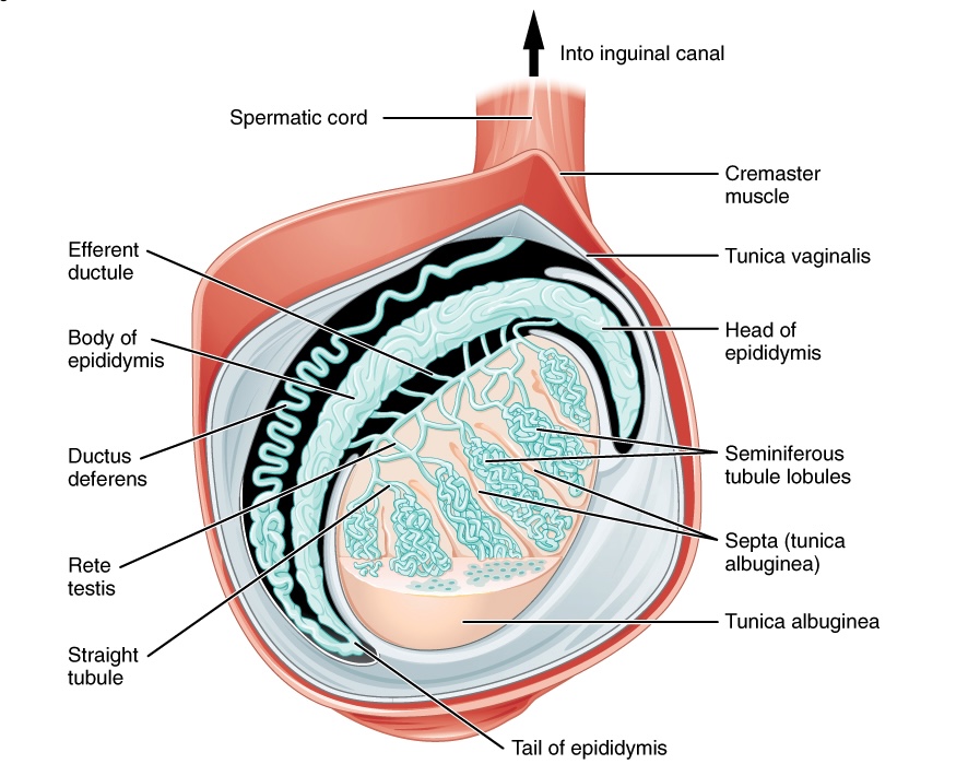
00:00 Now, we’re up towards the epididymis. On the right-hand side, you can see a section taken through the testis and the epididymis. Testis is shown here on the label, and then the epididymis is shown on the far right label. In between are the efferent ducts. Have a look at this histological section, including the testis, the efferent ducts, and the ductus epididymis. 00:33 And now, have a look at the figure. And just in your own mind, draw a line where you think the section passed when the histologist sliced the section through the testis, the efferent duct, and the epididymis to obtain the section that you’re looking at here. It is important sometimes in histology to be able to work out the orientation of how sections were actually taken through an organ. That’s just a little exercise for you to do partly of interest. Well, we now are looking at the epididymis. Have a look at the two sections shown here. Even though they are taken at two different magnifications, there are a few features I wish to point at. First of all, the bulk of the cells are called principal cells. 01:33 They exhibit stereocilia, long extended branching microvillus-like processes from the apex of their cells. They’re important to be able to absorb an enormous amount of fluid from the contents, from the lumen of the epididymis. Because the epididymis, one function is to absorb all the fluid that moves from the testis up into the epididymis duct, the fluid that’s added by secretions from the seminiferous tubule and by a secretion from the cells in the ducts I’ve already dealt with leading to the epididymis. 02:19 These stereocilia are then very important for that absorptive role. In the center of the lumen of each section through the epididymis shown here, you can see spermatozoa, many, many, many spermatozoa. The testis produces 300 million spermatozoa per day. Very different to the ovary, whereby, only one oocyte is ovulated every month. Huge contrast. 02:59 When you look at the epididymis, you can see smooth muscle around the wall, just underneath the lamina propria supporting the epithelium. On the left-hand side, you don’t see a lot of smooth muscles. On the right-hand side where the smooth muscle is labeled, you do. 03:19 And that’s because, on the left-hand section, you’re looking at a section through the epididymis taken towards the head of the epididymis, where really the prime role of the epididymis here is absorption. And therefore, you see a lot of stereocilia, as I’ve pointed out, on the apex of many other principal cells. Whereas, the section on the right contains a lot of smooth muscles. And that smooth muscle is important because, during ejaculation, that muscle is going to contract and force those spermatozoa along into the vas deferens which also contracts to expel the spermatozoa out through the penis during ejaculation. 04:09 So that muscle is very important and there are lots of layers of muscle there. The other thing you can’t notice really on this section, but I’ll tell you about it anyway, is that on the left-hand side, the epithelial cells that are doing all the absorption, they are very busy cells. They are tall. They are about 30 to 40 microns in height. Whereas, the ones on the right-hand side where the prime role is merely to move the spermatozoa into the vas deferens during ejaculation, these epithelial cells are half that size, half that height, and they don’t exhibit as many stereocilia on their surfaces.
About the Lecture
The lecture Epididymis – Male Reproductive System by Geoffrey Meyer, PhD is from the course Reproductive Histology.
Included Quiz Questions
Which of the following best describes the tail of the epididymis compared to the head?
- It contains the thinnest epithelium and the greatest quantity of smooth muscle.
- It contains the thickest epithelium and the greatest quantity of smooth muscle.
- It contains the thickest epithelium and the smallest quantity of smooth muscle.
- It contains the thinnest epithelium and the smallest quantity of smooth muscle.
The majority of the epithelium in the head of the epididymis contains which of the following types of cells?
- Columnar cells with stereocilia
- Simple cuboidal epithelium
- Clear cells
- Keratinized squamous cells
- Pyramid-shaped cells
Which of the following statements regarding the epididymis is INCORRECT?
- The stereocilia in the epididymis are motile.
- The stereocilia in the epididymis have a significant role in absorbing fluid.
- The head of the epididymis receives spermatozoa via the efferent ducts.
- During ejaculation, sperm flow from the epididymis into the vas deferens.
- The tail of the epididymis has the greatest quantity of smooth muscle.
Author of lecture Epididymis – Male Reproductive System

Geoffrey Meyer, PhD
Customer reviews
5,0 of 5 stars
| 5 Stars |
|
5 |
| 4 Stars |
|
0 |
| 3 Stars |
|
0 |
| 2 Stars |
|
0 |
| 1 Star |
|
0 |
