Playlist
Show Playlist
Hide Playlist
Enteroviruses – Picornaviruses

-
02-38 Picornaviruses.pdf
-
Download Lecture Overview
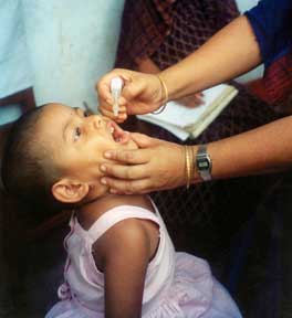
00:01 Let's start with the enteroviruses. 00:04 Now, the enterovirus family includes 3 major subcategories, which include enteroviruses, echoviruses, and coxsackieviruses. 00:14 Related to these 2 are the polio viruses. 00:18 The enteroviruses, as a whole, are resistant to acids such as one finds in the gastrointestinal tract. 00:24 They're also quite resistant to topical cleaning mechanisms such as detergent. 00:29 Transmission of all of these is fecal-oral. 00:33 Easy to remember because entero, gut, is the source of these viruses in human being. 00:39 Unfortunately, with robust exposure of vomitus or sometimes coughing or sneezing, there can be aerosol transmission as well. 00:49 Typical pathogenesis with the enteroviruses starts with a primary infection of the tissue target for that particular virus. 00:58 Many times, the initial infection starts in the pharynx, or the tonsils, the upper airway in lymphoid tissue, but then the viruses enter the gastrointestinal tract. 01:10 Where they spread, then, depends on the tissue tropism, and as mentioned, each virus may have a different specific target. 01:17 It is during that attachment to a specific tissue that the cytolytic replication occurs, meaning the cell is affected by or entered by the virus, the virus replicates itself, and when ready to leave with brand new virions, it destroys the cell. 01:35 That causes direct tissue damage, and then depending on the virus, the tissue damage could be anywhere. 01:42 In the case of polio virus, which we'll talk about next, the damage occurs at the level of the neurons and the neuro-motor junction, creating central nervous system disease. 01:53 For the other enteroviruses, coxsackie, echo, enteroviruses, broad tropism is noted. 01:59 Again, specifically viral subtypes may attack the brain causing central nervous system disease, the lungs, the heart, the pancreas, you name it. 02:09 So, let's look, then, specifically at poliomyelitis caused by polio virus. 02:16 And rarely, although this is not common, but rarely, coxsackie A virus. 02:21 Principally, polio virus is the cause of the disease known as poliomyelitis and it occurs in 3 different forms. 02:29 The first is what's known as abortive polio virus. 02:33 This is a very minor illness, which typically feels like a mild flu-like illness. 02:39 Low grade fevers, some malaise, headaches, basically the sort of thing which every one of us acquires several times a year and sleeps through for the next 3 days or so. 02:50 In some patients, this disease might actually be going into a nonparalytic form of poliomyelitis. 02:59 Of the patients who don't have the abortive virus, the remainder 80% will have this nonparalytic form. 03:06 Here, they start with that same viral process, viral syndrome, but it's accompanied by sore throat, sometimes developing into a stiff neck, and then they'll actually develop an aseptic meningitis. 03:19 Not severe in terms of prognosis, although it's very unpleasant with high fevers, severe headaches, a stiff neck, everything which would resemble a meningitis. 03:30 Of the 80% who go into a nonparalytic form, 10% actually may progress to a paralytic form. 03:38 So it is, sort of, the aseptic meningitis seen with nonparalytic, which then progresses with ongoing destruction of that lower motor neuron junction by action of the polio virus itself. 03:50 Those patients develop the flaccid paralysis, which we're all familiar with in looking at historical images, such as the iron lung. 03:59 So, moving on then to the enteroviruses, the non-polio enteroviruses, and the first disease category we'll talk about his herpangina. 04:09 Now, unlike the name, which sounds a whole awful lot like herpes, this has nothing to do with oral herpes or HSV type 1. 04:18 Instead, this is caused primarily by coxsackie A virus, sometimes by coxsackie B and other echoviruses. 04:25 But these viruses have tropism for, and among other things, the oral mucosa. 04:30 So a patient, typically a young patient, occasionally an older child to a young adult, will first develop fever and sore throat, along with some neck pain. 04:40 And then after several days of that, will develop incredibly painful lesions, ulcerative, or almost vesicular lesions on their posterior palate and over the tonsillar pillars. 04:52 And you can see a picture of this on the left side of the slide. 04:56 Those lesions are so painful that most patients find it impossible to swallow anything, ncluding their own secretions, and thus they'll present to health care because of the pain and dehydration. 05:09 Associated with that particular process is hand-foot-and-mouth diseases caused almost exclusively by coxsackie A virus. 05:18 These patients will have the same initial fever, sore throat, plus some nausea and vomiting, but then they will develop the same vesicular eruptions, not just in the back of the mouth such as herpangina, but also on the hands and the feet, typically, the palms and the soles. 05:36 Now, palmer and plantar rash should be a buzzword in your thinking because there are very few infections which can cause that. 05:43 Rocky Mountain spotted fever is one, secondary syphilis is the second, and coxsackie A virus causing hand- foot-and-mouth-diseases is a common third diseases causing palmer and plantar lesions. 05:56 These lesions are typically painful, so children will not wish to walk. 06:00 They'll be non-ambulatory. 06:02 Adults may experience pruritus, itching of the lesions, and it may be that children experience that also, but are not able to verbalize the feeling of pruritus. 06:14 Acute hemorrhagic conjunctivitis, also something that can be seen with coxsackie A virus. 06:20 And as you see in the picture, this is a very hemorrhagic inflammatory process affecting the conjunctival tissue, the entire conjunctival tissue, along with excessive tearing, mucus discharge, and even hemorrhaging of the subconjunctiva. 06:35 Again, you can see all these issues or all these elements on the picture in front of you. 06:40 This hemorrhagic conjunctivitis would be like an adenovirus, pink eye, gone really bad. 06:46 And in fact, adenovirus can do this too, but it's primarily the coxsackie viruses that have this severe appearance. 06:54 And then one of my favorites just because it sounds so neat, epidemic pleurodynia, which it has been named Bornholm diseases and even more -- and you'll love this -- it has been named devil's grip. 07:08 Grippe, G-R-I-P-P-E in historic language, and it has to do with how severely painful this is. 07:15 Coxsackie B virus causing a sudden, sharp paroxysmal chest pain, as if the devil has reached out of the grave and grabbed you by the chest. 07:25 That is how it used to be described, hence the name. 07:28 However, a Doctor Bornholm was the one who made it up bland and boring with the term epidemic pleurodynia. 07:35 In any event, these patients will have fever, severe headache, fatigue, muscle pain, typical viral syndrome, but with these sudden attacks of very sharp chest pain, which, in a way, limit breathing because they're so painful to take a deep breath. 07:49 Fortunately, the devil's grip is only 1 week long, resolves spontaneously, and one can only provide supportive care. 07:58 Aseptic meningitis. 08:00 Now, we're talking about the enteroviruses, including coxsackie and echoviruses that have tropism for the central nervous system. 08:07 These patients will start off with, again, fever, potentially a flat, erythematous, macular rash, which is nonspecific, and nausea, followed then by headache, neck pain, and the classic presentation of a meningitis. 08:22 Many of these will occur during the summer months or the warm climate months for the country of origin. 08:28 And patients will present and undergo evaluation for a bacterial meningitis. 08:34 They will be found to have a certain number of white blood cells in the spinal fluid, but with typically normal protein and glucose, suggesting that this is an aseptic or non-bacterial process. 08:48 And then neonatal disease. 08:50 Infants also can be born to mothers who are actively infected, typically with echo viruses and coxsackie B viruses. 08:57 And if so, they may be delivered with or soon develop after delivery signs of neonatal sepsis, which could be temperature instability, breathing failure requiring a ventilator, shock, hypotension requiring cardiac support. 09:15 Basically, depending on the virus and the organ-specific tropism of the virus, the babies could present somewhat differently. 09:22 The most common and most worrisome presentation of neonatal sepsis due to any congenitally or perinatally-acquired enterovirus is primary cardiac failure due to cardiomyopathy or myocarditis caused, again, by echoviruses. 09:40 So, how do I identify these antiviruses, the coxsackie echoviruses, coxsackie echos, the polio viruses? All these can be identified by looking at specific body tissues, including feces, blood serum, sometimes urine, sometimes respiratory specimens from a bronchoscopy. 10:01 And in taking a look at these specimens, one can look or for specific antibody, in fact, immunoglobulin M is the most acutely to be derived, although unfortunately the least sensitive in the laboratory. 10:16 You can look for a difference in acute versus convalescent immunoglobulin G antibody, expecting a rise of 4 fold or higher in the titer, but far more significantly and sensitively, we'll now use molecular diagnostics using a reverse transcriptase PCR to look at specimens of spinal fluid or blood to demonstrate that specific virus. 10:40 The picture you see in front of you is something we almost never do because that is a transmission electron microscope picture of an echo or actually enterovirus in the way that it looks, in clustered, beautifully symmetric form. 10:54 Yeah, we don't do that that often because, A, it takes forever and B, it's very expensive. 10:59 So, prevention and treatment. 11:01 In very few cases for the enteroviruses, or polio enterovirus, do we have the ability to prevent that, and polio virus is perhaps the best example. 11:11 There's been a long history of a mostly professional battle between doctors Salk and Sabin, who were at the same time almost in parallel trying to create a vaccine. 11:24 Now, history will suggest that Dr. Sabin's vaccine was the winner because his was the first that was produced in mass production. 11:31 It was an oral polio vaccine. 11:34 It was a live attenuated, meaning processed to avoid virulence vaccine, which was easy to administer and was administered worldwide. 11:44 Why not Dr. Salk's vaccine? Because the initial trials were poorly inactivated. 11:50 There was a problem with the inactivation process, and it actually was associated with some cases of secondary polio. 11:57 He went back to the drawing board and ultimately created a perfectly successful safe vaccine, which is in common used today. 12:05 What happened to make the difference occur between oral polio vaccine -- Sabin's product -- and injected or inactivated polio virus -- Salk's product? The challenge is that a live, attenuated virus still has some pathogenicity for those who are immunocompromised, and so, many patients in a family who were given the live oral polio vaccine from Sabin would shed the polio inactivated attenuated strain of virus, and somebody in the family who was immunocompromised would acquire that, fecal-oral transfer, would acquire that, fecal-oral transfer, So, due to safety reasons, the inactivated polio virus is what we use, ertainly in developed countries. 12:50 Although the oral polio vaccine because it is easy to administer and easy to deliver still used in many parts of third world countries. 12:59 Treatment for any of the enteroviruses can be attempted, but highly unsuccessfully with this medication called Pleconaril. 13:07 It's specifically has been tested at efficacy against enteroviruses, but it only works to mitigate the virulence of some of these some of the time. 13:16 So, tincture of time, lots of support, is the best way to go in treating any of these.
About the Lecture
The lecture Enteroviruses – Picornaviruses by Sean Elliott, MD is from the course Viruses.
Included Quiz Questions
Which of the following is considered the most likely viral etiology if a diagnosis of herpangina is established in a patient presenting with sore throat and painful vesiculopapular lesions on the soft palate?
- Coxsackie A virus
- Herpes simplex virus 1
- Herpes simplex virus 2
- Cytomegalovirus
- Reovirus
Sudden onset severe, paroxysmal chest pain associated with fever and headache caused by the Coxsackie B virus is known as...?
- ...epidemic pleurodynia.
- ...endemic pleurodynia.
- ...epidemic angina.
- ...endemic angina.
- ...endemic rachiodynia.
Which of the following refers to the management of choice for acute, severe chest pain occurring as a manifestation of Bornholm disease?
- Supportive care
- Intravenous acyclovir
- Oral famciclovir
- Oral acyclovir
- Intranasal pleconaril
Damage to which of the following structures by the poliovirus results in flaccid paralysis?
- Lower motor neurons
- Upper motor neurons
- Oligodendrocytes
- Schwann cells
- Satellite glial cells
Development of acute hemorrhagic conjunctivitis is usually a consequence of infection by which of the following enteroviruses?
- Coxsackie A virus
- Coxsackie B virus
- Echovirus
- Poliovirus
- Rhinovirus
Which of the following best describes the oral polio vaccine developed by Albert Sabin?
- Live-attenuated
- Whole-cell inactivated
- Conjugate
- Toxoid
- Subunit
Author of lecture Enteroviruses – Picornaviruses

Sean Elliott, MD
Customer reviews
5,0 of 5 stars
| 5 Stars |
|
5 |
| 4 Stars |
|
0 |
| 3 Stars |
|
0 |
| 2 Stars |
|
0 |
| 1 Star |
|
0 |
