Playlist
Show Playlist
Hide Playlist
Endoderm Derivatives and Gut Tube

-
Slides 02-07 Endoderm Derivatives and the Gut Tube.pdf
-
Reference List Embryology.pdf
-
Download Lecture Overview
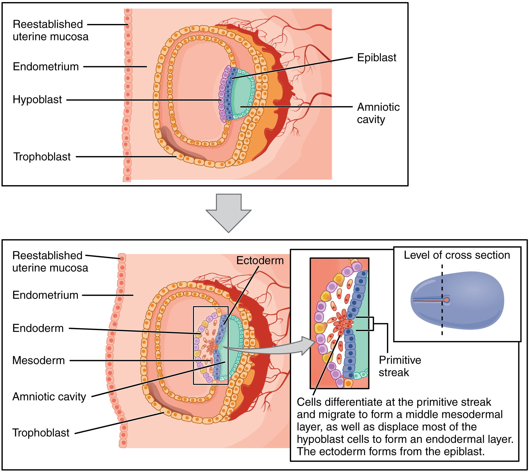
00:01 For our last talk on the derivatives of the trilaminar embryo, we´re gonna return and discuss how the endoderm forms the lining of the gastrointestinal, respiratory, and urogenital tracts. 00:12 Recall that it´s located far below the ectoderm and then the mesoderm and its development is tightly linked with the visceral layer of lateral plate mesoderm. 00:22 So the splanchnopleure which is the term we employ for endoderm and visceral layer of lateral plate mesoderm together is going to pinch together and form an actual tube. 00:33 Simultaneously, we´re gonna have the formation of large vessels just dorsal to it. 00:39 These are gonna be the paired dorsal aortae which are eventually going to fuse into a single aorta. 00:45 Now, as the somatopleure wraps around the body and creates the anterior body wall, simultaneously, the splanchnopleure and the endoderm that lines the inside of it pinched together to form a distinctive tube. 01:02 This is the gut tube and as it does so it separates from the yolk sac and the portion of the yolk sac that´s remaining is gonna now be called the secondary yolk sac. 01:12 Around day 25, we can also see that the intermediate mesoderm is forming the early kidney alongside the dorsal aortae. 01:23 The dorsal aortae are getting closer and closer and are fusing as this process occurs. 01:28 By the 26th day, the gut tube has completely come together and is connected to the dorsal body wall by what is called a dorsal mesentery. 01:38 Now, the dorsal mesentery is of vital importance because it´s the way that the blood vessels and innervation to the gut tube are actually able to reach it. 01:46 A byproduct of the folding is a ventral mesentery that connects the gut tube to the anterior body wall. 01:52 This disappears pretty much everywhere except the foregut and liver which is gonna be forming with something called the falciform ligament. 02:01 Now, the gut tube is connected to the anterior body wall because the somatopleure has come together and fused completely on the anterior aspect of the embryo and completely enclosed the intraembryonic coelom. 02:15 In the process, it has pinched the secondary yolk sac off of the gut tube and it only remains tethered to it by a small expansion from the midgut. 02:26 So if you look at this illustration, it would be easy to think the yolk sac is completely separate from the body but there is in fact one tiny point where it´s connected at what is gonna become our umbilicus. 02:37 So it may be appearing outside but it sticks around for a little while, but eventually, rescinds and doesn´t contribute anything to the mature developing embryo. 02:46 Before we move on, it´s important to get an idea about how the heart is actually tied to formation of the gut tube. 02:55 The heart does not actually come from the gut tube but it develops in close association to it. 02:59 You may recall that the cardiogenic mesoderm migrated very far anteriorly and in fact, our heart starts developing above our head and eventually, folds down into our chest. 03:11 There´s gonna be little hallow tubes that develop in the mesoderm called endocardial tubes and they come together and form a single endocardial tube as the body wall folds. 03:24 So here, we can take a look at this illustration and see the notochord right in the center. 03:30 Just below it is where we have the endoderm and the rest of the splanchnopleure folding together to create a gut tube. 03:37 As this folding occurs, the heart tubes on either side are moving anteriorly and are actually gonna take up residence just in front of the gut tube. 03:46 As they do so, they´re pulling a small space called the pericardial coelom along with them. 03:52 Now, as the folding of the splanchnopleure occurs, it brings these heart tubes closer together and actually allows them to fuse and we wind up with instead of two, just one endocardial tube that has the heart muscle develop around it. 04:07 Now, don´t worry if that´s a little unclear. 04:09 We´re gonna have an entire series of lectures on development of the heart. 04:12 But what I want you to note now is that the heart develops very much anterior or ventral to the foregut and is surrounded by a space called the pericardial cavity. 04:23 The heart has to come online and start beating by day 22 because at that point, the cavities that exist inside the embryo are no longer sufficient to allow diffusion of nutrients, waste products, and gasses, and we actually need to have a circulatory system develop and the heart being the pumping portion of that system is vitally important for a continued development of the embryo. 04:46 So here we can see that it´s remaining attached to the gut tube by a single dorsal mesocardium but one thing that you wanna appreciate about this picture is is that the heart´s maybe located ventral to the gut tube but it´s going to send vessels that go to every single organ of the body as development proceeds. 05:05 So as we move through this process, we come to a point where the embryo, instead of just being a collection of cells or stacked layers of cells is actually starting to look vaguely human. 05:15 So at day 22, we can see the neural tube folding together, day 24, we can see several somites laid out like beads on a string on either side of it. 05:25 And then, by day 26, if you squint, you might convince yourself you see some sort of animal there with the heart bulge sticking out in the front and the body wall beginning to fold around and completely enclose everything. 05:38 But here, as we move to day 28, we can see limb buds, distinctive head, a tail, which we eventually lose, and then, by day 32, once again, doesn´t look completely human but you can convince yourself that this is a developing animal with limb buds, heart, and head. 05:56 And we´re gonna return to the trilaminar embryos, the starting point for every lecture more or less moving on from here because all the organ systems can be followed from that trilaminar stage forward to make sense of it. 06:10 So thank you very much and we´ll return back here and move forward through every single organ system in the next series of lectures.
About the Lecture
The lecture Endoderm Derivatives and Gut Tube by Peter Ward, PhD is from the course Early Development and the Organogenic Period.
Included Quiz Questions
The dorsal mesentery, which suspends the gut tube, is a remnant of which embryonic layer?
- Lateral plate mesoderm
- Intermediate mesoderm
- Extraembryonic mesoderm
- Paraaxial mesoderm
- Cardiogenic mesoderm
The ventral mesentery disappears everywhere except at the liver during embryonic development and it persists after birth as what structure?
- Falciform ligament
- Round ligament
- Hepatoduodenal ligament
- Gastrosplenic ligament
- Triangular ligament
At what embryonic age does the heart usually begin to beat?
- Day 20–25
- Day 10–15
- Day 30–40
- Day 15–20
- Day 25–30
Author of lecture Endoderm Derivatives and Gut Tube

Peter Ward, PhD
Customer reviews
5,0 of 5 stars
| 5 Stars |
|
6 |
| 4 Stars |
|
0 |
| 3 Stars |
|
0 |
| 2 Stars |
|
0 |
| 1 Star |
|
0 |
This series of lectures saved me days of studying in my exam period in 1st semester of medschool :)
i like how he explain all and the images like support are amazing
thank you very much finally, i understood embryology you made it easy, understandable, and fun to learn
Great Teacher, enjoyed every single second watching these lectures. I couldn't ask for more. Great lectures Ever!
