Playlist
Show Playlist
Hide Playlist
Electrophoresis and Western Blot

-
17 Slides Immunodiagnostics.pdf
-
Reference List Immune System.pdf
-
Download Lecture Overview
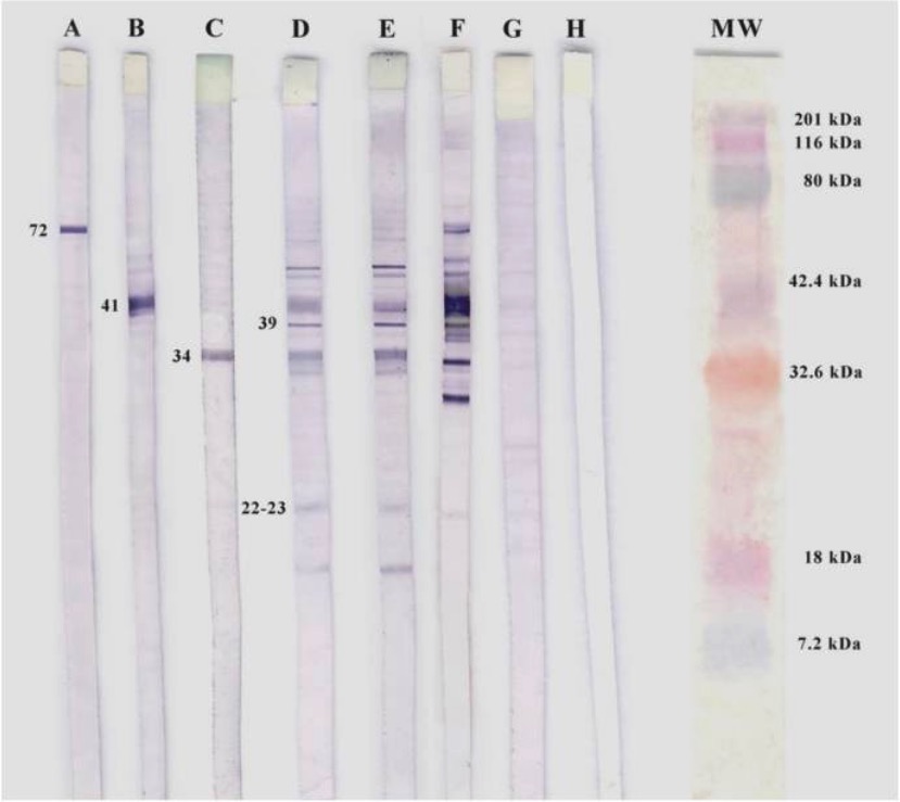
00:01 One can separate serum proteins using the technique of electrophoresis. 00:07 In this example, some blood is being collected from an individual. 00:13 The blood is allowed to clot and the cells are spun out from the blood. 00:18 And the serum is going to be analyzed using this method. 00:23 The serum is loaded onto a gel. 00:27 Under the influence of the electric field, proteins migrate to different regions in the gel. 00:33 The gel can then be stained to identify the location of the proteins. 00:38 Here we can see albumin, and also the different globulins that have migrated to different positions in the gel. 00:49 Immunoglobulins will migrate to the gamma globulin position. 00:58 Here’s an example of an electrophoretic analysis. 01:02 In the column A, a sample from a patient with a polyclonal expansion of B-cells, which has resulted in an increase in the gamma globulins. 01:16 Contrast that with the column B which is a normal control serum. 01:22 In column C, the sample is from a patient with a B-cell malignancy. 01:31 You can clearly see the gamma globulin from the dominant clone of B-cells in this patient. 01:38 Western blotting is a technique used for the analysis of proteins. 01:45 Here we can see a test tube with a mixture of protein antigens. 01:51 The proteins are denatured in the presence of SDS and applied to the gel. 01:59 Under the influence of an electric field, the proteins will be separated using this technique - SDS-poyacrylamide gel electrophoresis. 02:14 Following electrophoretic migration, the gel is removed from the tank and a membrane is placed next to the gel. 02:30 The buffer is added on filter papers, either side of the gel and membrane. 02:38 The cathode and anode plates are connected to a power supply. 02:45 And electrophoretic transfer of proteins from the gel to the membrane takes place. 02:52 Now one can label the different proteins on the membrane using a primary antibody specific for the antigen of interest. 03:03 To detect the binding of that antibody, a secondary antibody is added which is specific for the primary antibody. And this secondary antibody is tagged with an enzyme. A substrate is then added, for which the enzyme will cause an emission of light. So the substrate emits light when cleaved by the enzyme. This light is detected by autoradiography. And the membrane is exposed to a photographic film.
About the Lecture
The lecture Electrophoresis and Western Blot by Peter Delves, PhD is from the course Immunodiagnostics. It contains the following chapters:
- Separation of Serum Proteins by Electrophoresis
- Western Blotting
Included Quiz Questions
Which of the following regions of an electrophoresis contains the largest quantity of globulins?
- γ region
- α region
- β region
- Albumin region
- No region of an electrophoresis contains globulins.
In the western blot method, which of the following is generally used for the denaturing electrophoretic separation of proteins?
- Sodium dodecyl sulfate–polyacrylamide gel electrophoresis
- A polystyrene microtiter plate
- Immunoprecipitation
- Radial immunodiffusion
- Denatured albumin
After preparation of the membrane in western blot, which of the following are used as sensitive and specific detection tools that bind the protein antigen directly?
- Primary antibodies
- Secondary antibodies
- Enzymes
- Gels
- Anions and cations
Author of lecture Electrophoresis and Western Blot

Peter Delves, PhD
Customer reviews
5,0 of 5 stars
| 5 Stars |
|
1 |
| 4 Stars |
|
0 |
| 3 Stars |
|
0 |
| 2 Stars |
|
0 |
| 1 Star |
|
0 |
I understand it so well. Thank you for the lessons, professor.
