Playlist
Show Playlist
Hide Playlist
Ectoderm Derivatives and Neurulation

-
Slides 02-04 Ectoderm derivatives and neurulation.pdf
-
Reference List Embryology.pdf
-
Download Lecture Overview
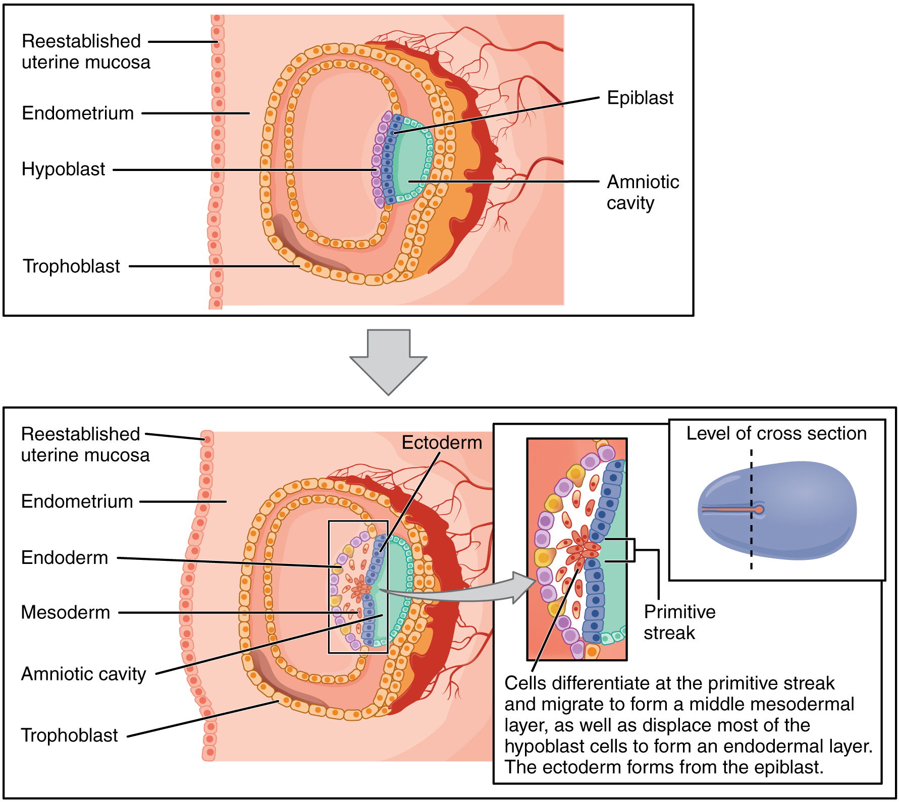
00:01 In this lecture we will follow how changes in the ectoderm are going to form the neural tube which will eventually become the entire central nervous system and its associated structures. 00:12 Now, when we left of in the prior talk we moved from the bilaminar embryo to the trilaminar embryo with ectoderm, mesoderm and endoderm. 00:21 Now, over the next several weeks, all of these tissues are gonna go through a great many changes to produce all the structures of the human body. 00:28 Now one thing to note is as we break these processes apart into further and further refine areas of the human development, all these processes are happening at the same time but to make sense of them it’s good to follow one organ system from start to more or less to finish before returning back to the others. 00:48 So to note, we’re gonna talk about the ectoderm folding to create the neural tube and central nervous system, as well as the epidermis of the skin. 00:57 But as this is happening, we have changes occurring in the mesoderm that will differentiate into somites which will form part of the musculoskeletal system. 01:06 The intermediate mesoderm will start turning into the urogenital system and the lateral plate mesoderm will become the supporting tissues of the gastroinstestinal tract as well as the body wall and limbs. 01:19 And last but not least, the endoderm will form a continuous tube called the gut tube which will become the lining of the gastrointestinal, urogenital and respiratory systems. 01:30 So let’s start with the trilaminar embryo and the notochord right in the center of the mesoderm. 01:37 It’s gonna drive changes that create paraxial mesoderm on either side of it, lateral mesoderm further out and intermediate mesoderm in between. 01:46 The endoderm which will form the gut tube is located just ventral to it and on its dorsal surface we have the ectoderm. 01:55 The ectoderm is going to thicken on either side flanking the notochord and there will be a nice little divot in between, it’s called the neural groove. 02:06 The neural groove will be a little bit thinner than the outside areas that are going to thicken to become the neural plate and the neural folds. 02:16 The neural folds are gonna grow upward and begin approaching one another on about day 20 of development. 02:23 And by the 21st day, we can see that these folds have not just grown closer and larger but they’re actually approaching one another almost ready to form a complete neural tube. 02:35 Now to see how this development proceeds a little further, we’re gonna focus simply on the neural tube and remove the mesoderm and endoderm from the picture. 02:44 In this illustration we can see the notochord in pink with the neural groove deepening closer into it as the neural groove migrates into the mesoderm. 02:54 On the dorsal side, the neural folds are approaching one another and moving into the mesoderm. 03:01 But as they do so, a group of cells called neural crest cells migrate into the mesoderm as well. 03:07 As development proceeds, the ectoderm on the outside or dorsal side of the body is going to fuse to form a completely seamless external skin which will become the epidermis. 03:22 The neural crest cells have migrated in to the mesoderm and will henceforth, begin migrating all over the body forming a variety of structures, in particular any nervous system ganglia located outside the central nervous system. 03:36 The neural tube is the major structure that results from the process of neurulation and it will become the entirety of the central nervous system from the spinal cord up to the brain. 03:47 The neural crest cells migrate in many directions, but one place they set up shop is on either side of the developing spinal cord where they will become the posterior root ganglia, sometimes also called the spinal ganglia. 04:01 We can now take a view of neurulation from the dorsal side. 04:06 So this is as though we’ve opened up the top of the developing embryo, we’re in the amniotic cavity looking down and we can see that the ectoderm is beginning to move into the mesoderm as neurulation proceeds and the neural tube forms. 04:19 On day 22, the process has begun and the neural tube is forming, moving into the mesoderm in about the midpoint of the embryo and then proceeds both caudally and cranially. 04:32 And so we have the neural tube moving in and zipping itself shut going forward and backwards simultaneously. 04:39 As this process proceeds, we’re left with an opening on the caudal end and the cranial end and eventually we have only one opening on the caudal side, the caudal neuropore, and on the cranial side, the cranial neuropore, marks the location of the eventual cerebral cortex. 04:58 If normal development proceeds, the cranial and caudal neuropores will also close, form a completely separate central nervous system and leave the amniotic space all together. 05:10 If this fails, a variety of birth defects can happen. 05:13 Chief among these with the cranial neural tube or the neuropore rather would be anencephaly. 05:20 If the cranial neuropore fails to close, the cerebral cortices cannot form that portion of central nervous system will not be present and the brain will not develop, hence, anencephaly, failure of the brain to form. 05:35 This can be detected by the levels of alpha-fetoprotein in the amnion. 05:40 Neural tube defects release alpha-fetoprotein in a level that can be detected with the maternal amnion sampling. However, this is rarely done. The current standard for screening for neural tubal defects is to measure the level of alpha-fetoprotein in the mother's serum and predict the risk based on the level of the serum alpha-fetoprotein and the gestational age. 06:05 If the caudal neuropore fails to close, the most important thing to recall is that you’ve got the central nervous system closing and only then can the skin close above it. 06:18 If we don’t have the caudal neuropore close on time, the vertebra cannot form around it on time and this results in a series of conditions called spina bifida and it can be more or less severe depending on how large and how late that neural tube defect sealed up. 06:38 The most innocent version of this is called spina bifida occulta or hidden spina bifida. 06:44 In this case, the neural tube did indeed close, did create a spinal cord and got surrounded by its supporting meninges, the arachnoid mater and the dura mater, but it closed late enough that the vertebra nearby are unable to meet on the midline and form a continuous lamina across the posterior or dorsal side of the spinal cord. 07:08 This is usually very clinically invisible, doesn’t really cause too many problems and may only be marked by a tuft of hair over the location of the neural tube defect. 07:17 A step up in the severity is if closure of the neuropore occurred much later and instead of having the skin evenly close around it, we’ve got a bubble and this bubble is called a meningocele and it includes cerebrospinal fluid and dura mater pushing out onto the skin. 07:39 This indicates that the neural tube closure was much later or much more significant than it would have been in spina bifida occulta. 07:45 A step further is called a meningomyelocele, indicating that both the meninges and the spinal cord have moved into this defect so the bubble doesn’t just contain CSF and meninges, it contains CSF, meninges, and nervous system tissue. 08:04 The most severe form of neural tube defect caudally is called myeloschisis or rachescisis. 08:11 In this case, the neural tube didn’t form and the neural plate never involuted and moved into the mesoderm so the presumptive nervous tissue is still open to the outside and is nonfunctional. 08:24 So any muscles caudal to this defect will be denervated and nonmobile. 08:29 Thank you very much for your attention and we will return and investigate other systems forming from the mesoderm and endoderm.
About the Lecture
The lecture Ectoderm Derivatives and Neurulation by Peter Ward, PhD is from the course Early Development and the Organogenic Period. It contains the following chapters:
- Changes in the Ectoderm which form the Neural Tube
- Failure of the Neural Folds
Included Quiz Questions
From which embryonic layer is the CNS derived?
- Ectoderm
- Intermediate mesoderm
- Lateral plate mesoderm
- Endoderm
- Paraaxial mesoderm
The organs of the urogenital system are derived from which embryonic layer?
- Intermediate mesoderm
- Ectoderm
- Lateral plate mesoderm
- Endoderm
- Paraaxial mesoderm
The neural ganglia arise from which embryonic structure?
- Neural crest
- Neural tube
- Neural canal
- Surface ectoderm
- Intermediate mesoderm
Failure in the cranial neuropore closure results in which of the following?
- Anencephaly
- Microcephaly
- Spina bifida
- Myeloschisis
- Meningocoele
A failure of the neural folds to close in time can prevent the vertebrae from forming properly but not cause displacement of nervous tissue or the dura mater. How would this congenital defect be classified?
- Spina bifida occulta
- Myeloschisis
- Meningocoele
- Anencephaly
- Microcephaly
Author of lecture Ectoderm Derivatives and Neurulation

Peter Ward, PhD
Customer reviews
5,0 of 5 stars
| 5 Stars |
|
4 |
| 4 Stars |
|
0 |
| 3 Stars |
|
0 |
| 2 Stars |
|
0 |
| 1 Star |
|
0 |
This video, with the previous two videos are amazing. Taking embryology in med school was like greek or hebrew. Your teaching style is very helpful to dissect the processes into understandable segments. Our professor taught by day of development so there was lots of jumping around. I like the discussion of one layer from flat layer to cells to folding, to closure, etc. Very well done. Moose Henderson, PhD
Clear and concise. Also appreciate the pace that he was going. Wish there were more questions though
It was easy for me to follow the professor as he was explaining the materials. I could understand the concepts and the process.
The lecture was clear and the material is high quality. I wish it was made a little bit clearer what the neural plate is.
