Playlist
Show Playlist
Hide Playlist
Dystrophin-associated Protein Complex

-
IRheumatology II 01 Muscle Pathology.pdf
-
Reference List Pathology.pdf
-
Download Lecture Overview
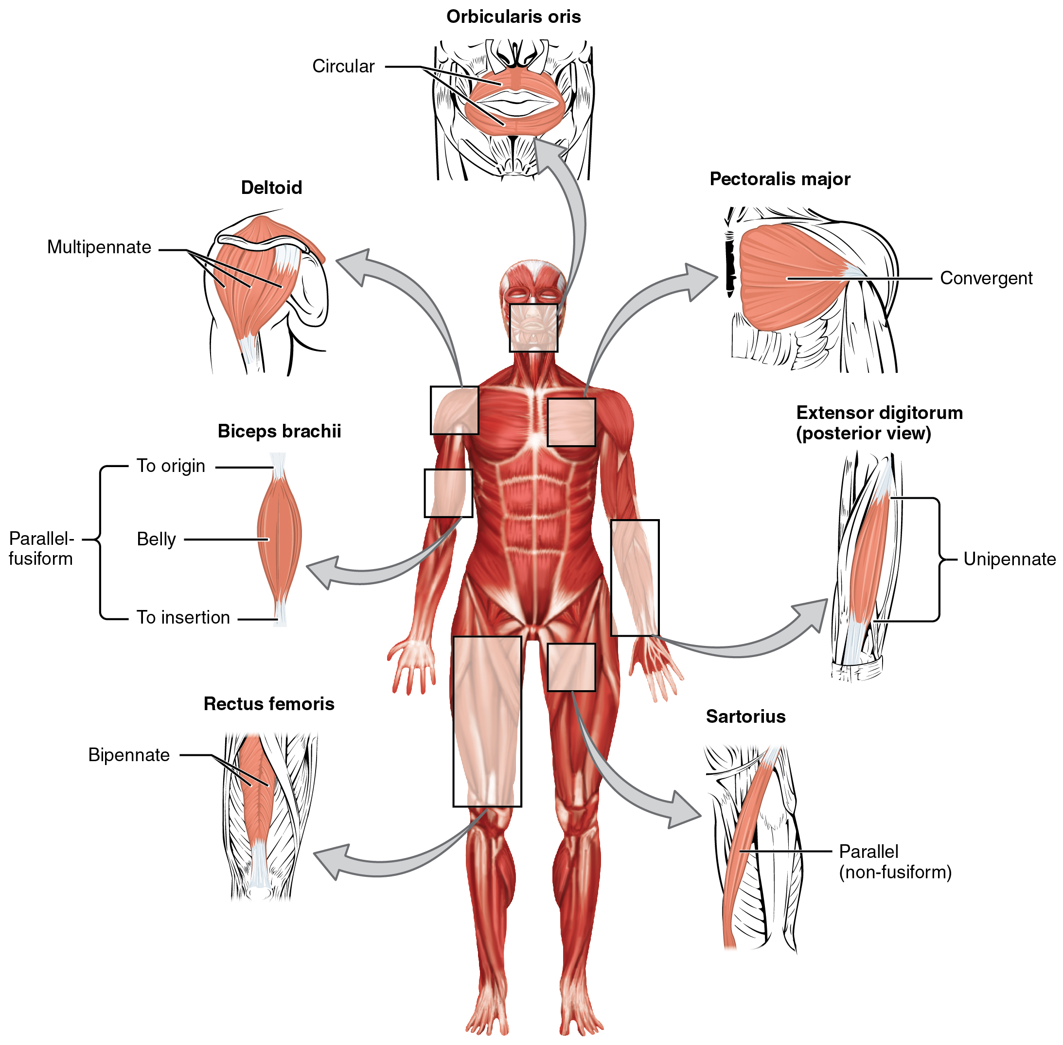
00:01 In this section, we’ll take a look at muscle pathology, and at first, I’m going to make sure that we walk through organization of a few things that I feel as though are important from anatomy and physiology that the pathology will make perfect sense. Let’s begin. 00:18 Classification of muscle itself—cardiac, of course, being involuntary. 00:24 Cardiac also then being striated intercalated discs so and so forth. 00:27 Keep that in mind. 00:29 With the cardiac muscle, the fundamental unit is going to be the sarcomere from Z line to Z line, and then you have the actin, and you have the myosin. 00:40 That is all intercellular and this then will communicate with the extracellular matrix and what may then happen, since we’re doing muscle pathology, are some of the anchors that are in the extracellular matrix to keep the intracellular components intact, might disappear. 01:01 Not only would it affect the cardiac muscle, but then it would also affect the skeletal muscle when we start talking about issues that we categorize as muscular dystrophies. 01:13 Smooth muscle also involuntary, but the smooth muscle is a little bit different, isn’t it? We don’t necessarily call it from Z line to Z line, we have these dense bodies. 01:24 Think about smooth muscle then you need to have relaxation, in other words, when a blood vessel relaxes, we’ll talk about vasodilation or if the bronchi, then you call it bronchodilation, and vice versa of course when it’s contracting in other words constriction. 01:41 The skeletal muscle, and the fundamental unit here would be similar to that of the cardiac and therefore when we comment to muscular dystrophies, our discussion will not only be relevant for the heart but then also for the skeletal muscle. 01:57 Quickly here, to review, our muscle and the types of muscle that you should b referring to when you see Z line to Z line, would then be your cardiac muscle and skeletal muscle, correct? What you’re seeing here on the left is electron microscopy. 02:13 It is important that you’re able to identify your sarcomere on electron microscopy. 02:20 You will then focus upon the Z line which is on the ends here and then the Z line will then have your titins and such without going into too much into anatomy and physio which will then bond your myosin to myosin represents a dark feature here. 02:36 You see the dark aspect of the dark band that you see here is your myosin. 02:41 And the light, of course, would then represent the actin. 02:45 And what you have there in the middle is the M line and you have the H band. 02:50 Now you must know of course that from Z line to Z line, when the length of it becomes shorter, obviously you’re in the process of contracting. And when you’re contracting, the A band will not change in length. 03:04 Because the A band will strictly then only be representing the myosin. 03:11 Whereas the I band, if you take a look at the I band, you have Z line to Z line, which will then contract and the I band and the H band will indeed change in length. 03:21 Keep that in mind. The one of the left is electron microscopy, once you see over to the right, focus on the A band please. 03:29 The A band is only representing that of the myosin; therefore, upon contraction. 03:37 The sarcomere terminology, while the actin is important that you know and this will then play a role as we begin pathology. 03:46 To review actin real quick, it is the light portion of the electron microscopy that I just showed you. 03:52 And the actin is the one that has the tropomyosins and the troponins. 03:57 Once calcium ________ it will untug the tropomyosins so that it can then bind to the myosin heads if you remember. 04:05 The A band will then represent the dark, in other words, only represents the myosin and therefore will not change in length upon contraction. 04:14 The H band was heavy chains only and the Z line from Z line represents the sarcomere. 04:22 The sarcomere terminology continues, the Z line things that change with contraction and the light bands shorten, and this then represents the I band. 04:32 Refer to the fact that it only represents the myosin and does not change in length upon contraction. 04:41 There are particular proteins that I want to make sure that you’re extremely familiar with and that your focus specifically is called to dystrophin. 04:49 Please note that dystrophin is the one of the largest proteins that is coded for in our bodies. 04:54 That would mean that if you have a protein that is highly coded, it would also mean that it’s susceptible to mutations. 05:02 Good. So when we later on have our discussion of muscular dystrophies it’s a fact that either have complete absence of your functional dystrophin or you might have partial retention of your dystrophin as we shall see. 05:19 Where is your dystrophin? Well once you hit your dystrophin let me set up this picture here—you have the membrane, the transmembrane, and we’ll be focusing upon those subunits specifically the beta and alpha. 05:30 They’re called your dystroglycans. 05:33 The alpha and beta dystroglycans, the transmembrane protein is the bridge between the dystrophin which is facing the extracellular matrix intercellularly intercellularly you will then have your, what we call, actin and company. 05:49 A combination of the two of after dystrophin, the anchor has been lost and you can imagine things that will take place and that will be a discussion later, but I want to make sure that you’re in or updated to what I need you to know for pathology. 06:05 Here, why do you want to know the different types of muscle fibers real quick? Well, it’s important. 06:11 For example, if you have a lady, less than 37 years of age and she wakes up in the morning and she feels okay, but then around let’s say 11 or 12 she works in front of a computer all day and she’s an assistant what have you, and she says that she’s having a hard time seeing, and now you ask yourself, what’s going on? And then later on at 4 o’clock maybe she has a hard time just moving, having a hard time just getting out of her chair. 06:36 So you do need to ask yourself of what kind of muscle fibers are being affected and why is the patient then having difficulty seeing at 11 or 12 o’clock The reason for that is the eyelids are your fast-twitch fibers—over to the right Type II muscle fibers are fast twitch. 06:51 The first type of muscle fiber to be affected in the lady with the type of diagnosis that she has is called myasthenia gravis. 07:01 Myasthenia gravis syndrome as we shall also discuss later, the eyelids which are, in general, the fast-twitch muscle fibers will be affected first. 07:11 Now before I move on, the opposite of your fast twitch obviously would be slow twitch, and a cute little mnemonic that I like using, and you can use whatever you wish, is I—Type 1—red, or RED or RED meaning aerobic glycolysis hemoglobin being supplied to that muscle slowly. 07:32 In other words, Type I muscle fiber is a red muscle fiber, , it is slow muscle fiber. 07:39 If it’s not Type I, it’s Type II. If it’s not red then it’s white. 07:43 If it’s not slow then it’s fast. Welcome to Type II. 07:48 Without going into further detail, I just want you to once again to have a comparative table to review between Type I and Type II without getting into too much physiology so that you’re clear and understanding that that you have Type II muscle fibers that are fast twitch, lots of ATPase activity, and so therefore can fatigue very, very quickly such as the eyelids; therefore, are one of the first type of muscle fibers that are affected in condition. 08:19 Now muscular dystrophies is what we look at first. 08:22 It’s a genetic issue most of them tend to be exocrine related. 08:27 Characteristically degeneration of not just skeletal muscle but then also cardiac muscle because of the similar, as I showed you earlier, microscopic features of Z line to Z line sarcomeres. 08:40 Now, because the muscles then are being broken down, obviously the enzyme that you’re expecting to then see to be elevated would be creatine kinase. 08:48 A differential, or how it’s differentiated, would be based on the age of the patient and when the onset of the disease was, what kind of muscles are involved, and what is the mode of inheritance is important. 09:02 Now you will have deletions that take place in both, and by both, we will be focusing on two major muscular dystrophies and then we will end by looking at myotonic dystrophy. 09:13 And as the order in which we will walk through muscular dystrophies, the nonspecific degenerative changes in biopsy include replacement of that muscle with what’s known as fibro fatty tissue. We then call this pseudofat. 09:28 Pseudofat. 09:30 So you have the, let’s say the, gastrocnemius or the calves in which it is then replaced by fibro fatty changes, in other words, pseudohypertrophy, is what it’s referred to. Pseudohypertrophy.
About the Lecture
The lecture Dystrophin-associated Protein Complex by Carlo Raj, MD is from the course Muscle and Soft Tissue: Pathology. It contains the following chapters:
- Introduction to Muscle Pathology
- Dystrophin-associated Protein Complex
- Lambert-Eaton Myasthenic Syndrome
Included Quiz Questions
What represents a sarcomere in skeletal muscle?
- It is the repeating unit between two Z lines.
- It is the repeating unit between two H zones.
- It is the repeating unit between two I bands.
- It is the repeating unit between 2 A bands.
- It is the repeating unit between a Z line and an I band.
The length of what structure remains the same during contraction?
- A band
- I band
- H zone
- Thin filaments
- Z line
Which one of the following components of the sarcomere is composed primarily of actin?
- I band
- Thick filaments
- H zone
- A band
- Z line
What is TRUE regarding type II fast-twitch muscle fibers?
- They are rich in ATPase.
- They are rich in myoglobin and mitochondria.
- They are found in the long muscles of the back.
- They do not fatigue easily.
- They are rich in oxidative enzymes.
Which of the following enzyme levels are elevated in muscular dystrophies?
- Creatine kinase
- Troponin T
- Troponin I
- Amylase
- Lipase
Author of lecture Dystrophin-associated Protein Complex

Carlo Raj, MD
Customer reviews
5,0 of 5 stars
| 5 Stars |
|
5 |
| 4 Stars |
|
0 |
| 3 Stars |
|
0 |
| 2 Stars |
|
0 |
| 1 Star |
|
0 |
