Playlist
Show Playlist
Hide Playlist
Dura Mater, Arachnoid Mater and Pia Mater
-
Slides 19 Meninges BrainAndNervousSystem.pdf
-
Reference List Anatomy.pdf
-
Download Lecture Overview
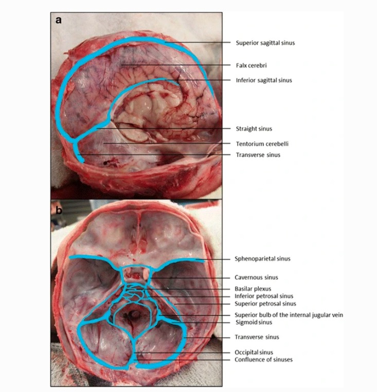
00:01 Now, let’s take a look at each of the three meningeal layers. The take home message here is for you to understand the basic characteristics of these meningeal layers. We’re going to start externally and work our way internally. So, that means we’ll look at the dura mater first and its characteristics. The dura mater is shown in green. In the cranial meningeal dura, the dura matter is going to be bilaminar. It will be a bilaminar, fibrous membrane. This is in contrast to the spinal dura mater which contains or is consisted of just one layer. Thus, it is unilaminar. So, if the cranial dura mater is bilaminar, what are the two layers that constitute the cranial dura mater? First is the outermost layer or the external most layer of the dura is going to be firmly attached to the inner part of the skull. That will be referred to as the periosteal layer. Then the internal or innermost lamina or layer of the dura will be the meningeal layer. These layers are fused with the exception of the dural venous sinuses where they will separate. That separation for example is shown here. This is the superior sagittal sinus. Right here attached to the inner part of the skull is the periosteal layer of the dura mater. Then this layer or lamina of the dura represents the meningeal layer. 01:58 Another area where they are not fused is where you have infoldings. So here, we have an infolding of the meningeal layers from either side of the skull. They are diving deep into the fissure that separates the two cerebral hemispheres. There are other examples of these types of infoldings that we’ll explore. So, what are the dural infoldings or reflections? We’ll walk through a few of them. One of them is not going to be represented on this particular slide. One will be represented in greater detail on a subsequent slide. But if we take a look at what we can appreciate in this slide, we have a major dural infolding. We see this infolding right in through here. This is the infolding that separates the cerebral hemispheres right from left. This is the falx cerebri. Another infolding and we see the cut edge of that infolding here and then we see the medial margin of it over in through here. 03:13 This is going to form a tent or a roof over the cerebellum. One of the cerebellar hemispheres rest down in this portion of the posterior cranial fossa and then the opposite hemisphere would rest or reside in the opposite area of the skull. Then the tentorium cerebelli will form a roof or a tent over those cerebellar hemispheres. A third type of infolding is the falx cerebelli. This is not a very significant infolding but it would attach to the tentorium cerebelli in the midline and then dive down between the cerebellar hemispheres but only for a short distance, not shown here on this slide, however. The fourth example of a dural infolding is that of the diaphragma sellae. That is seen in detail in the next slide. 04:17 The diaphragma sellae which is shown here and on the opposite side over here forms a roof over the pituitary gland that we see here residing within the sella turcica. If we look in the midline region of the diaphragma sellae, we see an aperture or an opening for the passage of the infundibulum that we see extending superiorly in this view. This will connect to the diencephalon. The last consideration here is the relationship of the diaphragma sellae with the pituitary gland as it pertains to a pituitary gland tumor. This diaphragma sellae is fairly fibrous, tough, so it does provide for resistance. So that if a tumor is forming, it’s less likely to move upwards, though it can. It’s more likely to take a path of least resistance and the tumor will expand laterally within the cavernous sinuses that we see here. 05:32 Now, let’s take a look at the arachnoid mater or simply arachnoid and its features. 05:40 The arachnoid mater is a thin membrane compared to the dura mater. We see that membrane right in through here. If we come over here to the right side of the image, we will see the arachnoid mater extend into the superior sagittal sinus. When the arachnoid mater extends into these types of venous channels between the periosteal dura mater and the meningeal dura mater, these form arachnoid granulations which help to move cerebrospinal fluid into the venous channels. The arachnoid also will have trabeculae or extensions that extend into the subarachnoid space which is the space here and then attached to the pia mater that is adherent or lining the surface of the cerebrum. It is not shown. 06:50 These are not shown, however, in this particular image. Again, we already highlighted the arachnoid granulations. Again, these are dilatations or expansions of the arachnoid where you have venous drainage or sinuses. Another feature of the arachnoid is that it is avascular. Lastly, the arachnoid mater is simply held against the inner dura mater which would be the meningeal layer by the force of cerebrospinal fluid pressure. In other words, it’s not adherent to, it’s just pushed up by this pressure. The innermost meningeal layer is the pia mater. Some of its features would include the fact that this is the thinnest and the most delicate of the meningeal layers. Again, we see it here as this thin, green line. 07:54 It will follow the contours of the cerebral cortex, the sulci, and the gyri. So here it lies over a gyrus and here it penetrates and dives into a sulcus. The pia mater is adherent to the surface of the brain. When you visualize this, it will confer a shiny appearance to the surface of the brain.
About the Lecture
The lecture Dura Mater, Arachnoid Mater and Pia Mater by Craig Canby, PhD is from the course Meninges. It contains the following chapters:
- Characteristics of the Dura Mater
- Characteristics of the Arachnoid Mater
- Characteristics of the Pia Mater
Included Quiz Questions
Which of the following statements regarding the dura mater is INCORRECT?
- The periosteal and meningeal layers are fused firmly with each other through the entire length of the dura mater.
- Cranial dura mater is bilaminar.
- The meningeal layer is the inner layer of the cranial dura mater.
- The periosteal layer is attached to the skull.
- Spinal dura mater is unilaminar.
At which point are the periosteal and meningeal layers of the dura mater distinctively separated?
- Dural venous sinuses
- The junction of the pia mater and arachnoid mater
- Outpouching of the dura mater
- Suture lines
- The junction of the dura and pia mater
Which of the following is NOT a feature of the arachnoid mater?
- It is a highly vascularized layer.
- It is thinner than the dura mater.
- It is held against the inner dura by CSF pressure.
- Trabeculae extend from the arachnoid mater into the subarachnoid space.
- It contains arachnoid granulations.
What is the term for the parts of the arachnoid membrane that project into the dural sinuses?
- Arachnoid granulations
- Confluence of sinuses
- Arachnoid cisterns
- Choroid plexus
- Arachnoid trabeculae
Which of the following statements regarding the pia mater is true?
- It confers a shiny appearance to the brain surface.
- It is responsible for the absorption of CSF.
- It is responsible for the production of CSF.
- It is the thickest of all layers of the meninges.
- It is not directly adherent to the brain surface.
Customer reviews
4,0 of 5 stars
| 5 Stars |
|
0 |
| 4 Stars |
|
1 |
| 3 Stars |
|
0 |
| 2 Stars |
|
0 |
| 1 Star |
|
0 |
The organisation of the layers of the meninges and outer regions of the cranium were well put together, but the diagram regarding in the infoldings (falx cerebrum...etc) was poor. I could find way better diagrams with a simple search onto google


