Playlist
Show Playlist
Hide Playlist
Duodenal Atresia in Children
-
Slides Structural GI Diseases.pdf
-
Download Lecture Overview
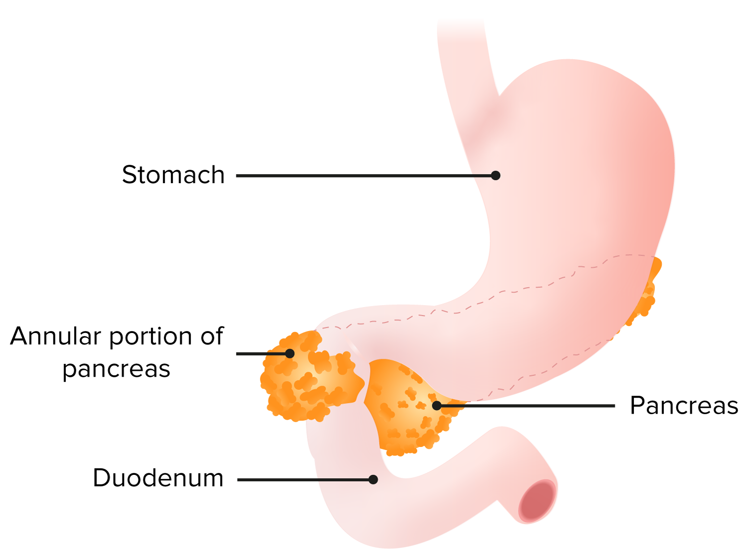
00:02 Moving on to duodenal atresia. 00:06 So here’s a classic case, a newborn with Down’s syndrome and abdominal distention. 00:12 This four-day old infant with known Down’s syndrome presents with abdominal distension suspicious of obstruction. 00:19 A child is vomiting just about everything he gets. 00:23 Clearly, Down’s syndrome and abdominal distention is a risk factor for duodenal atresia. 00:31 So intestinal atresias, all of them, are basically a failure of the hollow viscous organ to develop properly. 00:40 Instead of being hollow, it’s constricted and obstructs passage of food. 00:45 There’s a spectrum of severity, some are very badly atretic and some are only minimally atretic. 00:52 But what we do know is duodenal atresia is very closely associated with trisomy 21 or Down’s syndrome. 00:58 That is a very high-yield point on an exam. 01:01 Remember that connection. 01:03 So typically, the stenosis of any type of intestine, which is more common in the duodenum, occurs between two segments of bowel with or without a separating web. 01:16 The atresia will be a connecting fibrous band between those two segments of bowel and it can result in two blind pouches of bile, that’s the most common kind. 01:29 So what do we do in terms of diagnostic testing? Typically, we can get abdominal radiographs. 01:36 They will show obstruction, maybe pneumoperitoneum if it’s gone very far, and perhaps meconium peritonitis if it’s been left going too long. 01:46 The classic finding that is likely to be on your test is the double bubble sign. 01:52 What you will see is two round air-filled locations on an intestinal X-ray. 01:59 This is a picture of colonic atresia, not the double bubble sign, but the idea is still there. 02:05 You have an area where fluid is getting and then where fluid is not getting. 02:10 Sometimes, we’ll do contrast studies such as in this image, where an upper GI series may reveal a malrotation in addition to the double bubble. 02:20 In this case, where a patient has a microcolon, you can see an area where the colon is atretic. 02:28 Remember, and this is also high-yield for your exam, microcolon is associated with maternal gestational diabetes. 02:37 So duodenal atresia, Down’s syndrome, microcolon, maternal gestational diabetes. 02:45 Here is the double bubble sign. 02:47 And you can see the stomach filled with air and the duodenum filled with air as well. 02:53 The reason why there’s a line there is these are largely fluid contents because the child is presumably eating breast milk or formula, so it layers out in a gravitational manner. 03:04 Notice there is no gas distally. 03:06 This child is incapable of creating gas distally because nothing is getting into the distal intestinal compartment. 03:13 It is impossible to miss this for more than a few days because the child will pass away unless this is surgically corrected. 03:19 Food is not going through. 03:23 The surgical resection is essentially that. 03:26 They take out the atretic bowel and they put the healthy pieces of bowel back together as an anastomosis. 03:32 This allows the baby to recover and have full function.
About the Lecture
The lecture Duodenal Atresia in Children by Brian Alverson, MD is from the course Pediatric Gastroenterology.
Included Quiz Questions
What is a classic finding in a patient with duodenal atresia?
- 'Double bubble' sign
- Diarrhea
- Rash
- Difficulty breathing
- Urinary retention
Which of the following is a risk factor for duodenal atresia in children?
- Down syndrome
- Alpert syndrome
- Treacher Collins syndrome
- Cleidocranial dysplasia
- Hutchinson syndrome
Customer reviews
4,0 of 5 stars
| 5 Stars |
|
0 |
| 4 Stars |
|
1 |
| 3 Stars |
|
0 |
| 2 Stars |
|
0 |
| 1 Star |
|
0 |
Good lecture. All the necessary information for primary care resident is there. Thanks!


