Playlist
Show Playlist
Hide Playlist
Duct System – Male Reproductive System

-
Slides 07 Human Organ Systems Meyer.pdf
-
Reference List Histology.pdf
-
Download Lecture Overview
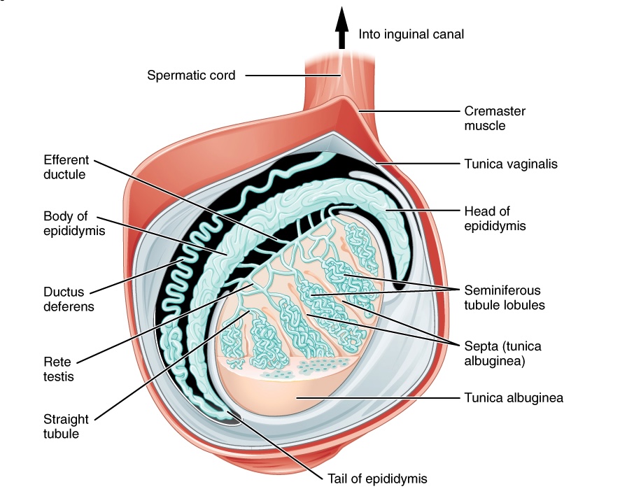
00:01 In this slide, you can see an illustration of the duct system that takes the spermatozoa away from the testis. And I’ve listed each of these ducts on the right-hand side and numbered them because I’m going to refer to these numbers to describe the duct system. Firstly, look at the testis. I’ve already described the testis in another lecture. But the testis has a capsule around it called the tunica albuginea. And that capsule divides the testis into a number of lobules. And these testicular lobules contain one to four long coiled tubes we call seminiferous tubule. And each of these tubules can be up to 50 centimeters long. So this is its enormous length of epithelium that is part of spermatogenic epithelium. This tube was aligned by seminiferous epithelium, which is the epithelium giving rise to the spermatogenic cells or containing the spermatogenic cells. And the final product of this epithelium is to release spermatozoa into the lumen of these tubules which then move their way towards the mediastinum of the testis. Before they get into the mediastinum of the testis, they pass into straight tubules. You can see them illustrated in the diagram and labelled 1 here. Those straight tubules then carry the spermatozoa along with fluid, into the mediastinum of the testis, into channels referred to as the rete testis, shown here labelled 2. And then, the fluid and the spermatozoa travel from the mediastinum into the epididymis via the efferent ducts. These efferent ducts are about 12 to 20 of them, they link the testis to the epididymis, the ductus epididymis. When you look at the epididymis, which is labelled 4 here, it’s a very long coiled tube. It can range from four to about six meters in length. It’s the site where sperm are going to go through their final maturation phase and become motile. It’s also the site that they’re stored. 02:53 Now, that epididymis has a head, a body, and a tail. The head is the component, the coil part of the epididymis right up next to the testis, joining the testis via these efferent ducts. The body is in the bulk of the epididymis passing around the posterior aspect of the testis. And then it starts to one coil at the tail region and it’s continuous then with the vas deferens labelled here, 5. I want you to keep in your minds a memory of this particular picture, because I’m going to refer now to all the different parts of the epididymis, the vas deferens and even all the duct systems coming out of the testis. 03:46 And I’m going to describe their histological features or characteristics and their importance. 03:55 Now on the right-hand side, you can see a histological section. This is taken right near where the seminiferous tubules become continuous with the straight tubule, number 1 in the diagram. You can see the seminiferous tubule, the seminiferous epithelium is minimal. 04:17 It’s not as thick as you see in other parts of the testis. It doesn’t contain a vast number of spermatogenic cells because the epithelium is changing as it moves from the seminiferous tubule to the straight tubule. In the straight tubule, it’s becoming cuboidal or even columnar, there're a variety of shapes of the epithelial cells, but generally speaking, they adopt the cuboidal type of appearance. And then the epithelium is maintained as we pass through the rete testis shown here on the right-hand section, and it’s equivalent to this section area on the diagram number 2. These are just huge channels or spaces that collect all the fluid containing the spermatozoa from the seminiferous tubules. 05:16 And they have a cuboidal, sometimes an even squamous epithelium. There is a little bit of a variety in these tubes. It’s particularly not that important, nor are the straight tubules in terms of defining their histology and their epithelial characteristics. The important point is that they are channels that are going to carry the spermatozoa from the testis up towards the epididymis. And then the efferent tubules look rather strange. They’re labelled 3 on the diagram, and they’re shown here in the histological section. Have a look at the epithelium. It’s rather an uneven surface, an undulating or corrugated type lumen due to the difference in the types of epithelial cells here. Some are cuboidal, some are columnar. 06:17 It looks as though some are even pseudostratified. It doesn’t matter too much. We tend to just term this particular tube a pseudostratified epithelium because really when you look at a number of sections through these tubes, the bulk of the epithelia illustrates a pseudostratified appearance. They have, on their surface, cilia. You can see some ciliated cells in this image. 06:49 And those cilia help to move the fluid along and carry therefore the spermatozoa into the epididymis. In the center of the lumen, you can see some red stained components that represent spermatozoa being transported along these ducts, the efferent ducts.
About the Lecture
The lecture Duct System – Male Reproductive System by Geoffrey Meyer, PhD is from the course Reproductive Histology.
Included Quiz Questions
Which of the following is located within the mediastinum of the testis?
- Rete testis
- Straight tubule
- Efferent duct
- Head of the epididymis
- Seminiferous tubules
What is the capsule of the testis called?
- Tunica albuginea
- Tunica vaginalis
- Tunica vuscularis
- Tunica testis
- Tubuli recti
Which of the following connects the rete testis to the epididymis?
- Efferent ductules
- Seminiferous tubules
- Seminal vesicles
- Vas deferens
- Tubuli seminiferi recti
What is the correct path from where the spermatozoa are produced to where they enter the urethra?
- Seminiferous tubules->rete testes->efferent duct->epididymis->vas deferens
- Rete testes->seminiferous tubules->efferent ducts->vas deferens->epididymis
- Vas deferens->epididymis->efferent ducts->rete testes->seminiferous tubules
- Seminiferous tubules->rete testes->epididymis->efferent duct->vas deferens
- Vas deferens->rete testes->epididymis->seminiferous tubules->efferent ducts
Which of the following is NOT a feature of the epididymis?
- It is where spermatozoa are initially produced.
- It has a head, body and tail.
- It is approximately 6 meters in length.
- It is where sperm is stored.
- It is where spermatozoa mature.
Author of lecture Duct System – Male Reproductive System

Geoffrey Meyer, PhD
Customer reviews
5,0 of 5 stars
| 5 Stars |
|
1 |
| 4 Stars |
|
0 |
| 3 Stars |
|
0 |
| 2 Stars |
|
0 |
| 1 Star |
|
0 |
Very informative. Brief, to the point. Clear explanation to facilitate understanding.
