Playlist
Show Playlist
Hide Playlist
Dorsal Aspect of the Hand

-
Slide Dorsal Aspect.pdf
-
Reference List Anatomy.pdf
-
Download Lecture Overview
00:01 In this lecture, we'll look at the anatomy of the hand continuing the series on the upper limb. 00:09 We'll start off by looking at the dorsum of the hand and the structures that are positioned within this area. 00:15 So, we can see the dorsum of the hand here of a right hand and we can see a number of structures passing from the forearm, the muscular structures that give rise to these tendinous structures that pass all the way through the hand into the distal aspect around the phalanges of the fingers. 00:33 If we just highlight some of these structures, we can then see the extensor digitorum. 00:38 We mentioned this in the forearm lecture on the posterior compartment and you can see that it extends alongside extensor indicis all the way to the phalanges of the fingers. 00:50 Here, we've got highlighted one muscle extensor, digiti minimi and this is specifically associated with the little finger. 00:57 But all of these tendons that pass from the forearm into the hand sit within the extensor retinaculum which is a thin band of tissue that helps to hold all of these tendons in place. 01:09 To orientate ourselves, we can see we have proximally, the carpal bones. 01:13 Then, we have a series of metacarpals and sitting around the digits where these tendinous insertions are passing into are a series of what are called extensor hoods and these are really important structures as they help to keep all of these tendinous insertions organized and help a nice tight structure around each of the phalanges. 01:36 Connecting to the tendinous insertions that are passing away from all of these extensor tendons are a series of intertendinous connections. And really, what this does is this means that all of the fingers are extended in unison. It's really quite difficult to extend certain fingers at one time because of all of these intertendinous connections. 01:58 You can do it around the index finger and the little finger because they have specific muscle bellies associated with those digits on their own. 02:07 But really, these intertendinous connections allow all of the fingers to work in unison and distribute some of the force related with that extensor movement. 02:17 So, let's have a look at these extensor hoods and what's within them in a little bit more detail. 02:21 So, here, we can see the extensor tendon is passing all the way through to the middle and the distal phalanx. 02:28 Here, we can see the medial tract. So, just the medial portion of this extensor hood is passing towards this middle phalanx which we can see here. 02:37 Whereas you have these two lateral tracts that run either side of that medial tract and that passes toward the distal phalanx which we can see here. 02:45 And these, like I said, help to distribute all of the force of those extensor muscles to help when you're extending your fingers. 02:52 Here, we can see the extensor expansion which is really an elevated covering of this tight connective tissue. 02:59 And we can see how it's roughly triangular in shape. But importantly, it has this free margin which allows those tendons to pass in and then, associate with the middle and distal phalanges. 03:12 The triangular shape of this extensor expansion covers the dorsal sides of the phalanges. 03:17 So, both the lateral and the medial sides of these phalanges to provide this tight covering around those digits. 03:26 Also inserting into this space are the intrinsic muscles of the hand. 03:30 And that helps them to work with their function which we'll come to later on. 03:35 Another important space within the hand is what's known as the anatomical snuff box. 03:40 So, the anatomical snuff box is indicated here in these green dotted lines. 03:45 And we can see it's got a base. It's also got an apex. It's broadly triangular in shape. 03:51 It has a medial border which is formed by extensor pollicis longus tendon. 03:57 So, the tendon of that muscle. And then, two muscles form its lateral border. 04:02 We've got extensor pollicis brevis and abductor pollicis longus. 04:07 So, we can see the anatomical snuff box is really a space that's created by the position of these series of tendons. 04:14 It's an important space to be aware of because deep within it are two important bones, the scaphoid bone and the trapezium. These two bones form the floor of the anatomical snuff box. 04:27 Also passing through, entering from the lateral aspect, we've got the radial artery and we also have branches coming from the radial nerve. 04:37 Sitting quite proximally within this space, we have the styloid process of the radius and more distally towards the apex, we have the first metacarpal bone, that of the thumb that sits distally. 04:50 This space that we've just described is important because during palpation, if pain is radiating from this space, it may indicate that there's been a fracture of the scaphoid bone. 05:01 And this bone is quite prone to being fracturing. 05:04 So, there's an important examination to take place and knowing where to find the anatomical snuff box is important. 05:10 So, they are the boundaries of that space. 05:13 Another important structure within the dorsum of the hand is the extensor retinaculum. 05:18 We mentioned that previously in this tight band that goes around the dorsal aspect of the hand. 05:23 And that helps to keep all of these tendons in position. Deep to it are a series of tendinous sheaths. 05:29 And these are really synovial sheaths that reduce friction as the extensor tendons are passing between this space as they're extending the fingers and as they're moving as the fingers are being flexed. 05:43 Also sitting on the dorsum of the hand, we have some superficial veins. 05:47 We can see this dorsal network of venous structures which are really draining all of the venous blood from the fingers and from the metacarpals and from the carpals. 05:56 And they will ultimately drain into even the cephalic or the basilic veins as we mentioned in the subcutaneous vasculature lecture. 06:05 We mentioned the tendons of extensor muscles. We can see them here. 06:08 And running just deep to those are a whole series of blood vessels around the carpal and metacarpal bones. 06:14 And these are supplying the structures on the dorsal surface, things like the interossei muscles which we'll come to later on. And this is the dorsal carpal arterial arch. 06:24 So, it's an arterial arch that runs across the carpal bones and it's on the dorsal surface. 06:29 So, the dorsal carpal arterial arch. And like I said here, it's supplying those interosseous muscles.
About the Lecture
The lecture Dorsal Aspect of the Hand by James Pickering, PhD is from the course Anatomy of the Hand.
Included Quiz Questions
Which statements about the anatomical snuff box are correct? Select all that apply.
- Its medial border is formed by the extensor pollicis longus.
- Its lateral border is formed by the tendon of extensor indicis.
- Its lateral border is formed by the extensor pollicis brevis.
- Its lateral border is formed by the tendon of abductor pollicis longus.
- Its floor is formed by the scaphoid and trapezium.
What is the extensor retinaculum?
- Fibrous band that holds the extensor tendons
- Ring that holds the extensor tendons
- Ring that holds the flexor tendons
- Fibrous band that holds the flexor tendons
- Flexor muscle
What helps the fingers function as one unit?
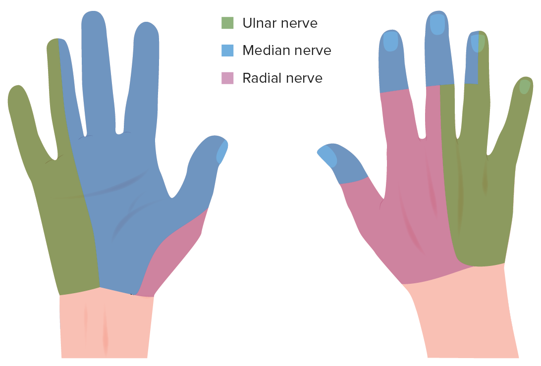
- Intertendinous connections
- Extensor retinaculum
- Extensor tendons
- Flexor hoods
- Flexor tendons
What is the shape of the extensor expansion?
- Triangular
- Rectangular
- Cuboidal
- Longitudinal
- Lateral
Author of lecture Dorsal Aspect of the Hand

James Pickering, PhD
Customer reviews
5,0 of 5 stars
| 5 Stars |
|
1 |
| 4 Stars |
|
0 |
| 3 Stars |
|
0 |
| 2 Stars |
|
0 |
| 1 Star |
|
0 |
This lecture is short and clear, making it easier to even remember the anatomical terms for the bones.
