Playlist
Show Playlist
Hide Playlist
Differences between Small Arteries and Small Venules

-
Slides 02 Human Organ Systems Meyer.pdf
-
Reference List Histology.pdf
-
Download Lecture Overview
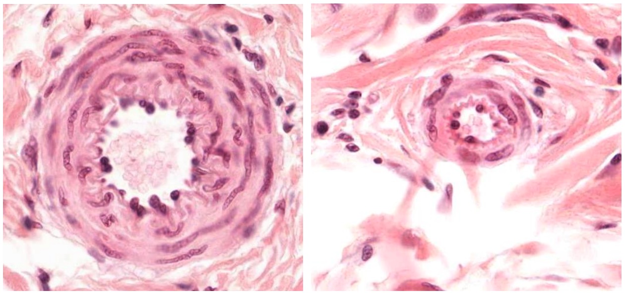
00:01 Well, let’s have a look at the structure of vein, a small artery and a small venule, and a postcapillary venule. On the bottom left-hand side of the image, the section on the left-hand side and on the bottom of the left-hand side of the image on the right-hand side, is a section through a small artery. It’s nice and circular. It has got a thick wall of smooth muscle. 00:30 But have a look at the vessel on the left-hand image on the top right, large lumen, much thinner wall. That’s the small vein. On the right-hand section down the bottom, again, large lumen, relatively small wall. That again is a small vein. So that's how you can tell the difference between a small artery and a small vein closely next to each other or in the tissue. And again, look at the dimensions, the relative thickness of the walls of these two vessels relative to the lumen diameter. Up on top of the image on the right-hand side is a large lumen or space. You can actually see blood cells within this lumen and a very, very very thin wall. This is a very small venule. It’s called a postcapillary venule. 01:30 Often, small venules and postcapillary venules are very different or very difficult rather to distinguish. But these very small venules are the end part of the capillary bed. 01:45 They’re receiving blood that has gone through all the capillary beds and that blood is accumulating in these venules. And then that will finally move on to the larger venules and larger veins as they move towards the heart. So make sure you’re going to understand how to distinguish the structures between a small artery, a small vein or venule, and then also the postcapillary venules. I think if you look at the right-hand image again and down the bottom, that small structure with a very thin wall is probably a small venule, whereas above, is a postcapillary venule. But really, the distinction isn’t that critical. It’s just to make sure you have an understanding that blood flows down through an artery, goes into a capillary bed which has very, very thin walls to allow diffusion, and we’ll talk about those in a moment, and then that blood passes into smaller veins, smaller venules, and then postcapillary venules, and finally back into larger veins and back into the cardiovascular system to return that blood back to the heart.
About the Lecture
The lecture Differences between Small Arteries and Small Venules by Geoffrey Meyer, PhD is from the course Cardiovascular Histology.
Included Quiz Questions
Which of the following statements regarding arterioles and/or venules is CORRECT?
- Arterioles appear circular on cross section.
- Venules have a smaller diameter.
- Venules have thicker walls.
- Venules have more layers of smooth muscle.
- Venules hold a lesser proportion of blood.
Which of the following types of vessels immediately follows the capillary bed?
- Post-capillary venule
- Vein
- Artery
- Arteriole
- Lymph vessels
Author of lecture Differences between Small Arteries and Small Venules

Geoffrey Meyer, PhD
Customer reviews
5,0 of 5 stars
| 5 Stars |
|
5 |
| 4 Stars |
|
0 |
| 3 Stars |
|
0 |
| 2 Stars |
|
0 |
| 1 Star |
|
0 |
