Playlist
Show Playlist
Hide Playlist
Development of Sutures and Fontanelles


-
Slides 04-15 Growth of the Skull.pdf
-
Reference List Embryology.pdf
-
Download Lecture Overview
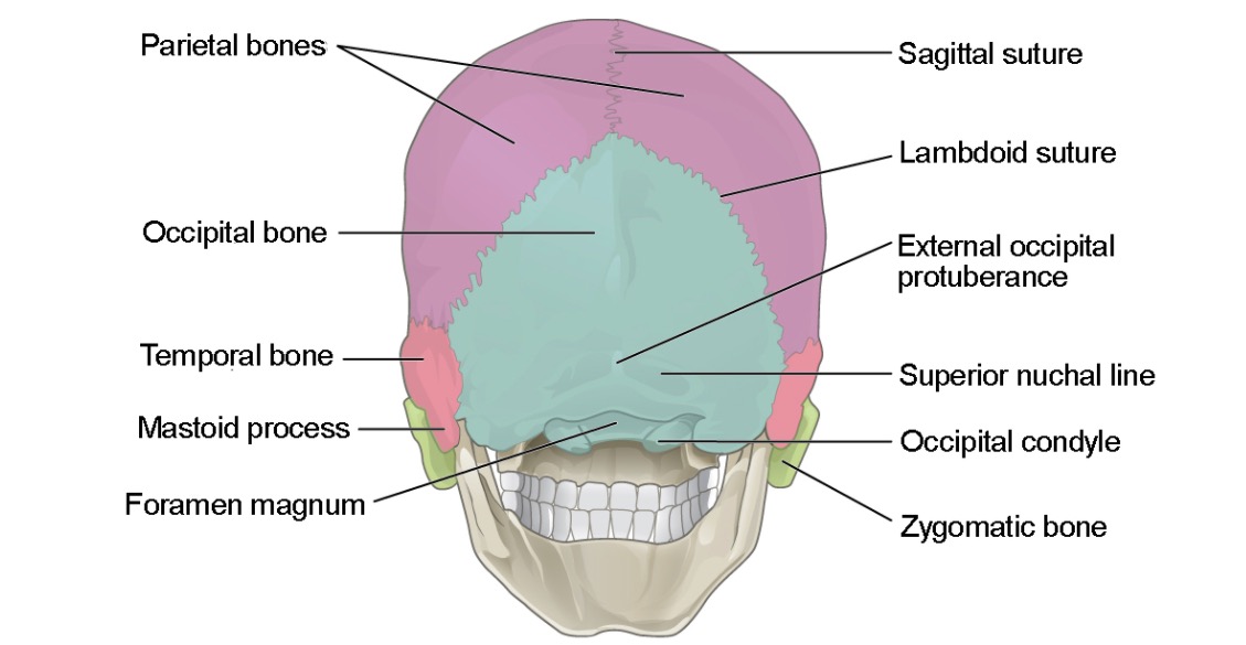
00:01 The sutures and fontanelles that are present in an infant´s skull are there because these bones are not allowed to actually grow completely together. 00:11 Instead of fusing, connective tissue between them forms and the more motion and more stress that these bones are exposed to during development creates more or less complex sutures between those bones. 00:24 So here´s a side view of the skull of an infant. 00:29 We can see that the bones are present but there´s actual separation between them and their neighbors. 00:36 Let´s review some of the bony features of the skull. 00:39 But before we move on, note here that the parietal and frontal bones have a very pronounced eminence. 00:46 Let´s take a look at it from above and here we can see that the parietal bones are sticking out, as are the frontal bones. 00:53 That´s where that initial core of ossification started. 00:57 As we get older, those eminences rescind a little bit and smooth out along with the rest of the bone. 01:04 So here between the two frontal bones is what´s called the frontal or metopic suture. 01:11 Now, this suture closes very early and most people aren´t aware that they have one. 01:15 We´re used to seeing a single frontal bone on the forehead. 01:18 But around 15% of the population still maintains this frontal suture and if you ever feel a pronounced bump right down your forehead, congratulations, you may be one of the lucky few. 01:28 Just posterior to that where that frontal suture meets the next one, the coronal suture is where we find the anterior fontanelle. 01:37 You are almost certainly more familiar with this as the soft spot that´s present in the head of an infant or a newborn. 01:44 So this soft spot is there because the bones have not yet fused. 01:48 The parietal and frontal bones have not grown together and because of that, there´s a spot that´s literally soft and if there´s extra pressure inside the brain, you can actually detect bulging of that soft spot clinically. 02:02 Now, if we move a little further down the coronal suture between a frontal and a parietal bone, we come to the anterolateral fontanelle where the temporal bone, the sphenoid bone, the frontal bone, and the parietal bones all meet. 02:20 In the adult, that little spot is gonna be called the pterion but early on, it´s a place where those bones meet and have not yet fused. 02:28 So a small anterolateral fontanelle and not surprisingly, there´s also gonna be a posterolateral fontanelle. 02:34 This fontanelle is gonna be found between the parietal bone, the occipital bone, and the temporal bone and again, it´s very small but it is present early on. 02:44 Between the two parietal bones, we find the sagittal suture going right down the midline of the skull. 02:52 Now the sagittal suture is still pretty pronounced in almost everyone until very extended old age. 02:59 Anterior to that between the parietal bones and the frontal bones is the coronal suture right here. 03:06 Sits like a tiara, coronal means crown, so it sits right there on our skull. 03:11 And very far posteriorly between the two parietal bones and the occipital bone is our lambdoid suture. 03:18 This image clearly shows the squamous suture not seen in the prior image. 03:23 That´s present between the temporal bone and the parietal bone. 03:27 So it´s located right about here. 03:29 And last but not least, there´s a small posterior fontanelle. 03:33 You find it at the juncture of the occipital bone and the two parietal bones. 03:39 So the thing to remember about those fontanelles is that there are four present. 03:43 Actually, I should say six present because we have a posterior lateral and anterolateral on the left and the right, and on the midline, you have a posterior fontanelle and a very large anterior fontanelle. 03:56 Now, one byproduct of the fact that we have endochondral ossification occurring in our skull is that our skull and our face are growing. 04:04 That´s not too surprising but if that entire new set of bone was solid, we´d have a lot of wasted space. 04:12 We´d have a lot of dense bone taking up space, making metabolic demands on us that we don´t really need. 04:18 For that reason, we develop paranasal sinuses. 04:21 These sinuses are air sacs connected to our nasal cavity that develop within the bone. 04:28 And they´re there because we don´t need bone in those places to have structural support for our skull. 04:34 So there are several well characterized nasal sinuses. 04:38 The frontal and a little bit posterior to that, multiple ethmoidal air cells. 04:43 In the cheek, the maxillary sinus, and very far back right at the base of the pituitary gland is where we find the sphenoid or sphenoidal sinus. 04:53 Now, these sinuses develop as the skull enlarges and that endochondral ossification creates more space than the skull needs for densely packed bone. 05:04 But most of these are not present until the skull has a change to enlarge. 05:08 So they´re not present at birth but enlarge as we age and the older we get, the more extensive and more space these sinuses take up. 05:17 Now, the maxillary sinus in the cheek is in fact present before we´re born. 05:22 It´s the only one that´s universally pretty much acknowledged to be there prior to birth. 05:27 Next, in the ethmoid bone, we have ethmoid air cells. 05:31 They´re present within two years of birth. 05:33 Thereafter, the sphenoid sinus begins forming around the same time but our frontal sinus really isn´t very early to the game. 05:41 It comes along about seven years later and not every child is gonna be able to get a sinus infection until they´re old enough to have those sinuses.
About the Lecture
The lecture Development of Sutures and Fontanelles by Peter Ward, PhD is from the course Development of the Nervous System, Head, and Neck.
Included Quiz Questions
What suture separates the left and right frontal bones?
- Metopic suture
- Coronal suture
- Lambdoid suture
- Squamosal suture
- Sagittal suture
The large anterior fontanelle is formed from the conjunction of the coronal suture and what other suture?
- Sagittal suture
- Frontal suture
- Squamosal suture
- Lambdoid suture
- Sphenofrontal suture
Which paranasal sinus is present before birth?
- Maxillary sinus
- Anterior ethmoidal sinus
- Posterior ethmoidal sinus
- Frontal sinus
- Sphenoid sinus
Author of lecture Development of Sutures and Fontanelles

Peter Ward, PhD
Customer reviews
5,0 of 5 stars
| 5 Stars |
|
5 |
| 4 Stars |
|
0 |
| 3 Stars |
|
0 |
| 2 Stars |
|
0 |
| 1 Star |
|
0 |
