Playlist
Show Playlist
Hide Playlist
Deep Layer (AC) – Anatomy of the Forearm

-
Slides 06 UpperLimbAnatomy Pickering.pdf
-
Download Lecture Overview
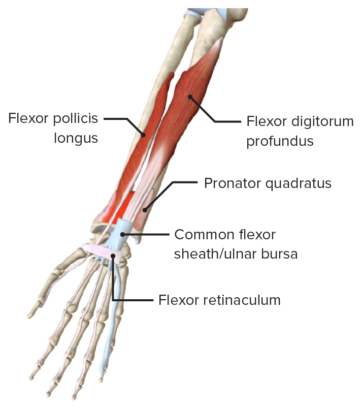
00:00 If we then look at the deep layer, we can see we've got some muscles which are lying deep to flexor digitorum superficialis. 00:09 We have flexor digitorum profundus. 00:13 We can see that this is coming from the ulna. 00:16 It's also coming from the interosseous membrane that's between the two bones of the forearm, the radius and the ulna. 00:24 And this again gives rise to four long tendons that this time go towards the distal phalanx of each digit, two to five, not including the thumb. 00:36 The thumb is different because it has its own specific flexure and this is known as flexor pollicis longus. 00:43 Pollicis meaning the thumb. 00:45 Flexor pollicis longus. 00:47 The fact that we call it flexor pollicis longus indicates is going to be a flexor pollicis brevis. 00:54 And this muscle is within the hands. So we'll look at it later. 00:57 The final muscle, the deep muscle in the anterior compartment is pronator quadratus. 01:03 And that runs between the two distal ends of the radius and the ulna. 01:08 So let's have a look at the attachment of these muscles. 01:11 Flexor digitorum profundus is running from the proximal surfaces of the medial and anterior surfaces of ulna, and the interosseous membrane. 01:22 So it's coming from the ulna. 01:25 It then passes via its four long tendons to the distal phalanges, the medial four distal phalanges of digits 2, 3, 4, and 5. 01:38 The nerve supply, it has a jewel nerve supply. 01:42 The lateral muscle bellies, these the ones that are going to digits two, and three are supplied by the anterior interosseous nerve. 01:50 And the tendons that are going to digits four and five are supplied by the ulna nerve. 01:57 So flexor digitorum profundus has two nerves supplying it, the median nerve and the ulna nerve. 02:06 Its function is to flex the hand at the wrist joints, and it also flexes the distal interphalangeal joint. 02:13 So where flexor digitorum superficialis flex the proximal interphalangeal joint. 02:19 This knife flexes the very distal interphalangeal joint. 02:23 The joint between the distal phalanx and the middle phalanx. 02:28 If we go back and look at flexor pollicis longus, we can see this is really coming from the radius. 02:34 And this gives a long tendon that goes and attaches to the distal phalanx of the thumb. 02:41 So we can see that flexor pollicis longus running from the anterior surface of the radius, and also the interosseous membrane passes to the distal phalanx of the first digit. 02:51 First digit be in your thumb. 02:54 The pronator quadratus, as we saw here, is running between the two distal ends of the radius and the ulna. 03:01 It runs from the distal quarter of the ulna. 03:04 So the last quarter of the ulna and it actually passes towards the radius. 03:10 So when pronator quadratus contracts, it is actually going to move the radius. 03:15 It is going to move the radius into a more pronated position. 03:18 So from this supine position, contraction of pronator quadratus is going to protonate the radius, pronate the forearm. 03:28 It pulls the radius over the ulna bone. 03:31 Both flexor pollicis longus and pronator quadratus are innervated by the anterior interosseous nerve. 03:40 And this is a branch of the median nerve. 03:42 So the median nerve is supplying the majority of these muscles within the anterior compartment. 03:48 But a specific branch the anterior interosseous nerve is supplying the flexor pollicis longus and pronator quadratus. 03:58 Flexor pollicis longus does similar functions to flexor digitorum muscles except it acts at the first digit. 04:06 Continued contraction will flex the wrist, it flexes the interphalangeal, and metacarpophalangeal joints associated with the first digit. 04:17 Pronator quadratus pronates the forearm. 04:20 So it acts on the radioulnar joints.
About the Lecture
The lecture Deep Layer (AC) – Anatomy of the Forearm by James Pickering, PhD is from the course Upper Limb Anatomy [Archive].
Included Quiz Questions
Which nerve innervates the lateral muscle bellies of the flexor digitorum profundus?
- Anterior interosseous nerve (branch of the median nerve)
- Ulnar nerve
- Brachial nerve
- Radial nerve
- Musculocutaneous nerve
Which movement is performed by the flexor digitorum profundus?
- Flexion of the distal interphalangeal joint
- Extension of the hand at the wrist joint
- Flexion of the proximal interphalangeal joint
- Extension of the metacarpophalangeal joint
- Extension of the distal interphalangeal joint
Author of lecture Deep Layer (AC) – Anatomy of the Forearm

James Pickering, PhD
Customer reviews
5,0 of 5 stars
| 5 Stars |
|
1 |
| 4 Stars |
|
0 |
| 3 Stars |
|
0 |
| 2 Stars |
|
0 |
| 1 Star |
|
0 |
Thank you for this video, loved how concise and precise it was
